Uncategorized
BBB Access Route via Proteomic Vascular Mapping
A limiting feature of many neurological therapies is the ability of molecules to cross the blood-brain barrier (BBB) from the circulatory system. Since the BBB prevents simple diffusion of materials across the divide, identifying the proteins responsible for transport is necessary for effective design of BBB-crossing therapies.
“So basically, everything in the circulating blood, if they want to have an exchange with the organ, they need to pass through this interface,” says senior author Jiefu Li, PhD, Janelia Research Campus Group Leader at the Howard Hughes Medical Institute.
Identification of the structures within blood vessels involved with the processes of molecular movement across the BBB has been somewhat elusive. However, Li and his team have developed a technique that not only identifies proteins within the luminal surface—the inner lining—of the vasculature, but also works in vivo, allowing them to track how these features change across the aging brain.
“Understanding how the blood-brain barrier works, particularly figuring out the molecular targets that you can play with to open and close the barrier, will provide new possibilities for drug delivery,” Li says.
Their work is published in Science in a paper entitled, “Luminal surface proteome of the brain vasculature uncovers blood-brain barrier regulators.”
Using mice, the team developed a proteomic profiling method that can be used not only in brain vasculature, but throughout the body. “Briefly, a lectin-conjugated peroxidase is perfused and anchored to the luminal surface of blood vessels to catalyze the biotinylation of adjacent proteins, thereby enabling subsequent protein enrichment and mass spectrometry analysis,” wrote the authors.
They tested the method in the brain, kidney and intestine, in both mice and northern tree shrew, showing functionality and applicability across organs and species.
“This will allow us to say: we know that the vasculature system is doing different things in different organs and it relies on this luminal surface, but how does that happen? What are the molecular players there?” Li says.
Using quantitative proteomics of the luminal surface—from early development through adulthood and aging—they found that over time there was a decrease in angiogenic and transport proteins. They also found an increase in proteins that increased stiffness in the vasculature.
In addition to developing this in vivo technique, the team identified two proteins that are temporally distinct in their expression while both playing a role in modulating BBB permeability. Knockouts of nitric oxide synthase Nos3 and arginine transporter Slc7a1 resulted in BBB leakage in neonates, but not adults, while genetic screens identified hyaluronidase HYAL2 as being required for maintaining BBB integrity throughout the lifespan of mice.
“What we know now is that we have two new pathways, potentially, to open the blood-brain barrier and to inform some therapeutic developments,” says Li.
Utilization of this proteomic based method in vivo both opens up new avenues of functional research across the cardiovascular system, and also provides data and a methodology for novel therapeutic targets for crossing the BBB.
“This method solves an important need but it’s also a very easy-to-use method, so everyone can use it,” Li says.
The post BBB Access Route via Proteomic Vascular Mapping appeared first on GEN – Genetic Engineering and Biotechnology News.
Uncategorized
STAT+: At hospital finance conference, a call to end the friction that’s keeping costs high
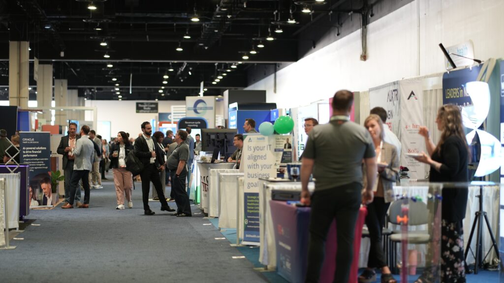
NATIONAL HARBOR, Md. — At this week’s annual meeting of hospital finance leaders, the exhibit hall was packed with dozens of billing and collections companies. Armed with candy, tote bags, and pens, they smiled at passersby, eager to explain why their tactics would extract the most money from health insurers.
The sheer number of “revenue cycle” vendors who attended the Healthcare Financial Management Association’s annual conference in Maryland — outnumbering even the hospital attendees, according to a list shared by an organizer — was a visible reminder of the enormous industry built around just paying medical bills.
The U.S. health care industry spends roughly $200 billion annually on financial transactions: claims processing, payment, collections, and prior authorization. And yet the proliferation of billing vendors seemed to clash with the main theme of HFMA’s conference, affordability, spotlighting the need to simplify the billing process so that health care is less costly and more accessible for patients.
Strong science, lower costs and growing capital networks are putting Spain and Portugal on the biotech investment map, even as structural bottlenecks persist, according to two investors.

You know when you are at the eye doctor getting an updated prescription, and suddenly the world snaps into sharper focus? Physicists at the University of California (UC), Berkeley, have now done something similar for electron microscopy. By introducing phase contrast into a cryo‑electron microscope, they have delivered dramatically sharper images of some of biology’s smallest and most elusive proteins.
The advance comes from a new laser phase plate (LPP), described in the paper “Laser phase plate improves structure determination of small proteins by cryo‑EM,” which was published recently in Science. Led by physicist Holger Mueller, PhD, of UC Berkeley and Lawrence Berkeley National Laboratory, the team demonstrated that a laser‑driven phase plate can overcome one of cryo‑EM’s most persistent limitations: poor contrast for small proteins.

Cryo‑EM has transformed structural biology over the past decade, earning a Nobel Prize in 2017 for enabling high‑resolution structures without crystallization. But despite its impact, the technique still struggles with proteins below ~70 kilodaltons—a size range that includes about 90% of the human proteome. “Because of signal-to-noise limitations, the majority of human and animal proteins are too small to be analyzed by these methods [cryo-EM and cryoelectron tomography]. The increase in signal-to-noise ratio provided by this laser phase plate is expected to overcome these important limitations.”
The new LPP begins to address that problem. The LPP uses an intense, continuous‑wave laser to shift the phase of the electron beam itself. This produces true phase contrast without dimming or destabilizing the beam. Mueller described the laser focus as “75 kilowatts focused to a few microns… That’s more powerful than what you use for welding. It has more power than a military laser. It builds up the brightest continuous laser focus ever.”
Installed in a custom Thermo Fisher Titan Krios, the LPP immediately improved the clarity and resolvability of small proteins, including hemoglobin, which sits at the lower limit of what today’s cryo‑EM instruments can handle. As the authors wrote in the abstract: “Here, we show that the laser phase plate (LPP)… enhances the resolution in single-particle reconstruction of small proteins by improving specimen-motion correction, recovery of information from the early frames, as well as particle visualization, 3D classification, and alignment.”

These improvements were achieved using standard defocus ranges and reconstruction workflows. “For the most challenging cases—small particles, bad specimens—the laser produces a very considerable advantage,” Mueller said.
The impact extends beyond single‑particle analysis. Cryo‑electron tomography (cryo‑ET), which assembles multiple angular views of a molecule or protein into a three-dimensional image, stands to benefit even more. “With cryo-ET, we’re looking at small, very complicated cellular material that’s incredibly crowded inside the cell,” said Bridget Carragher, PhD, founding technical director of imaging at Biohub. “It’s like a forest of trees, and you’re trying to find one leaf on one tree in there. Cryo-ET needs a dramatic step forward in contrast, so we can start to see what’s going on inside the cell. That’s what the laser phase plate promises to give us.”
Biohub is developing a dual‑laser version of the system, designed to reduce component wear and minimize aberrations. Meanwhile, Mueller’s team is pushing toward imaging proteins as small as 17 kilodaltons, a threshold that would open access to vast regions of the human proteome previously invisible to cryo‑EM.
“This technology is a step function change for biology,” said Stephani Otte, PhD, Biohub’s vice president of imaging science. “What was once invisible will become visible—and that changes everything about how we understand disease.”
“The bottom line is, if you have a large protein and a really good sample—a fresh one or one frozen without bubbles, for example—you may not need the phase plate to get a single, high-quality image. But for a small protein and a bad sample, laser-on is best,” Mueller said. “This could fill an enormous gap in our knowledge of protein structures that can’t be crystallized or are too small for today’s cryo-EM. And it will be revolutionary for cryo-ET.”
The post Laser‑Driven Phase Contrast Enhances Cryo‑EM Resolution of Small Proteins appeared first on GEN – Genetic Engineering and Biotechnology News.


Illinois’ financial crisis could bring the state to a halt
The final 6 ‘Game of Thrones’ episodes might feel like a full season
New Season 8 Walking Dead trailer flashes forward in time
Mod turns ‘Counter-Strike’ into a ‘Tekken’ clone with fighting chickens
Meet Superman’s grandfather in new trailer for Krypton
Disney’s live-action Aladdin finally finds its stars

STAT+: At hospital finance conference, a call to end the friction that’s keeping costs high
Beyond sunshine: Iberia’s biotech moment has arrived with developing capital networks

Laser‑Driven Phase Contrast Enhances Cryo‑EM Resolution of Small Proteins

STAT+: Updated: Tracking RFK Jr.’s promises to remake health in America

An obesity drug deep-dive, and peptides move mainstream

RFK Jr. claims his calendar is publicly available. We’ve been trying to get it for a year
Illinois’ financial crisis could bring the state to a halt
The final 6 ‘Game of Thrones’ episodes might feel like a full season
New Season 8 Walking Dead trailer flashes forward in time
Mod turns ‘Counter-Strike’ into a ‘Tekken’ clone with fighting chickens
Meet Superman’s grandfather in new trailer for Krypton
Disney’s live-action Aladdin finally finds its stars
-
 Uncategorized9 years ago
These ’90s fashion trends are making a comeback in 2017
-
Uncategorized9 years ago
According to Dior Couture, this taboo fashion accessory is back
-
Endpoints News3 months ago
Novartis to pay $2B upfront to take next-gen PI3Kα inhibitor from Synnovation
-
Uncategorized9 years ago
Phillies’ Aaron Altherr makes mind-boggling barehanded play
-
Uncategorized9 years ago
Uber and Lyft are finally available in all of New York State
-
Contributors9 years ago
The final 6 ‘Game of Thrones’ episodes might feel like a full season
-
Uncategorized9 years ago
Steph Curry finally got the contract he deserves from the Warriors
-
Uncategorized9 years ago
The old and New Edition cast comes together to perform