
Uncategorized
AI Uses MRIs to Generate Brain Aging Maps for Neurodegenerative Disease Research

Researchers at the University of Southern California have developed an approach that uses artificial intelligence to generate detailed maps that highlight differences in how distinct parts of the brain age. The researchers, led by associate professor Andrei Irimia, PhD, at the USC Leonard Davis School of Gerontology, used magnetic resonance imaging (MRI) from nearly 15,000 cognitively healthy individuals to train a deep learning AI model. The data provided a baseline against which the model could measure local brain age (LBA), or how old specific regions of the brain appear.
While most studies of brain age measure this phenomenon using a single number, the new model provides a much richer picture of typical aging and neurodegeneration. Rather than assigning a single “brain age” (BA) to an individual, the approach generates a detailed map showing how old different parts of the brain appear relative to what is typical for someone of the same chronological age.
When the AI model was then used to analyze MRI images from people with mild cognitive impairment and Alzheimer’s disease (AD), it revealed distinct patterns of accelerated aging in brain regions known to be affected early in neurodegeneration.
“Not all brain regions age at the same rate,” Irimia said. “Some areas appear to be more resilient, while others are more vulnerable to aging and disease. By measuring local brain aging, we can identify where the brain is aging faster than expected and how those changes relate to cognitive function.”
In their in paper in PNAS, titled “Deep learning maps local brain aging in relation to cognition across human adulthood,” senior author Irimia and colleagues stated, “By providing spatially resolved measures of brain aging, this work enables more precise investigation of how neuroanatomic alterations and cognitive impairment affect brain anatomy, above and beyond global brain age measures.”
Aging is a prominent risk factor for the onset of brain diseases, including Alzheimer’s disease and related dementias, the authors wrote. “One of the most prominent biological features of brain aging is atrophy, i.e., brain volume decrease that often involves loss of brain cells and neural connectivity.”
The newly reported research builds on previous efforts to estimate BA, an emerging neuroimaging biomarker that compares a person’s brain structure to patterns seen in healthy people across the lifespan. But while human brain aging is not uniform across cortical regions, traditional methods typically reduce the brain to a single age estimate, which can obscure important regional differences. The new approach instead measures local brain age at the voxel level—the three-dimensional units that make up an MRI scan—producing a much more detailed picture of structural aging throughout the brain.
“This more nuanced understanding of how the brain ages could pave the way for earlier identification of dementia, a better understanding of what factors affect risk and new ideas for treatment approaches,” Irimia said.
To develop the model, the researchers trained a deep-learning neural network using MRI scans from 14,748 cognitively normal adults ages 19 years to 100 years, drawn from six large public datasets, including the UK Biobank, the Human Connectome Project and the Alzheimer’s Disease Neuroimaging Initiative. The team then tested the model using MRI scans from more than 1,900 additional participants in the Alzheimer’s Disease Neuroimaging Initiative, including cognitively normal adults, people with mild cognitive impairment and people with Alzheimer’s disease.
Across healthy adults, the model consistently found that the frontal and temporal lobes—regions involved in decision-making, memory and other higher cognitive functions—appeared biologically older than the parietal and occipital regions, which are involved in spatial awareness and sensory processing functions. “Our approach consistently reveals spatial patterns of aging, including relatively advanced aging in frontal and temporal regions, across both typical aging and Alzheimer’s disease,” the investigators noted. The researchers also found that the brain’s right hemisphere tended to show slightly more advanced aging than the left, a pattern that persisted regardless of whether participants were right- or left-handed.
As cognitive impairment progressed, the differences became even more pronounced. Compared with cognitively normal adults, participants with mild cognitive impairment (MCI) or Alzheimer’s disease showed significantly older local brain ages in structures that are among the first affected by Alzheimer’s pathology, including the hippocampus, amygdala and several deep brain regions involved in memory and cognitive processing.
The researchers also found that older local brain age was associated with poorer performance on cognitive assessments, strengthening the link between structural brain changes and real-world function. “Deviations from normative regional aging are significantly associated with cognitive performance supported by neural processes linked to those regions … thereby relating anatomic aging to functional outcomes,” they stated. The strongest relationships appeared in people with Alzheimer’s disease, suggesting that regional brain aging may become increasingly informative as neurodegeneration advances.
Because the model produces anatomically detailed maps, it could eventually help scientists better understand why some people experience faster decline in specific cognitive abilities than others. The approach may also prove useful for tracking disease progression or evaluating whether experimental therapies are slowing degeneration in targeted brain regions. “By providing spatially resolved measures of brain aging, this work enables more precise investigation of how neuroanatomic alterations and cognitive impairment affect brain anatomy, above and beyond global brain age measures,” they commented.
Although the findings are promising, Irimia emphasized that the method remains a research tool. The model was trained primarily on research-quality MRI data and will require additional validation using more diverse clinical datasets before it can be adopted in routine patient care. The study also relied largely on cross-sectional data, meaning that future longitudinal studies will be needed to determine whether local brain aging can reliably predict who will progress from healthy aging to mild cognitive impairment or Alzheimer’s disease.
Still, the researchers believe that moving beyond a single measure of brain age represents an important advance for neuroscience. “By quantifying the anatomy of brain aging and aligning it with cognition and disease stage, this work establishes a foundation for mechanistic inquiry and personalized intervention in neurodegeneration,” the authors stated. “This scalable framework paves the way for monitoring a broad spectrum of neurodegenerative and aging related disorders,” Irimia added, “Brain aging isn’t uniform. “By understanding how individual regions age, as well as how those patterns differ from person to person, we’re moving toward a much more precise understanding of healthy aging and neurodegenerative disease. Ultimately, that could help us identify people at risk earlier and develop more personalized approaches to preserving brain health.”
The post AI Uses MRIs to Generate Brain Aging Maps for Neurodegenerative Disease Research appeared first on GEN – Genetic Engineering and Biotechnology News.

Uncategorized
STAT+: Takeda’s narcolepsy drug approved by the FDA, seen as a boon for new class of treatments
The Food and Drug Administration on Wednesday approved a novel type of treatment for narcolepsy made by Takeda, backing a drug class that scientists hope can transform the treatment of sleep disorders and potentially address a broad range of neurologic conditions.
The twice-a-day pill, which will be marketed as Orzeyful, is an orexin receptor agonist. It’s cleared to treat narcolepsy type 1, a taxing condition in which patients have a deficiency of the orexin neuron and experience bouts of sleepiness and muscle weakness during the day.
The drug is likely to be very appealing to patients. Current therapies, stimulants and sodium oxybates, carry risk of misuse, and patients still feel sleepy while taking them. In two Phase 3 trials, Orzeyful helped patients stay awake during the day for much longer than what’s been seen with current treatments. Throughout the studies, participants also reported less daytime sleepiness, less frequent muscle weakness, and greater attentiveness.

The Food and Drug Administration on Wednesday approved a novel type of treatment for narcolepsy made by Takeda, backing a drug class that scientists hope can transform the treatment of sleep disorders and potentially address a broad range of neurologic conditions.
The twice-a-day pill, which will be marketed as Orzeyful, is an orexin receptor agonist. It’s cleared to treat narcolepsy type 1, a taxing condition in which patients have a deficiency of the orexin neuron and experience bouts of sleepiness and muscle weakness during the day.
The drug is likely to be very appealing to patients. Current therapies, stimulants and sodium oxybates, carry risk of misuse, and patients still feel sleepy while taking them. In two Phase 3 trials, Orzeyful helped patients stay awake during the day for much longer than what’s been seen with current treatments. Throughout the studies, participants also reported less daytime sleepiness, less frequent muscle weakness, and greater attentiveness.
Uncategorized
Erica Schwartz confirmed as CDC director, filling nearly yearlong vacancy
WASHINGTON — Erica Schwartz was confirmed Wednesday as the director of the Centers for Disease Control and Prevention, making her the second permanent leader of the agency since the second Trump administration took office.
Her confirmation, approved by the Senate in a 51-44 vote, also ends a nearly yearlong vacancy for the CDC’s top job after health secretary Robert F. Kennedy Jr. fired Schwartz’s predecessor last year.
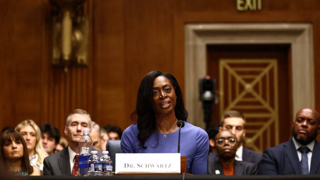
WASHINGTON — Erica Schwartz was confirmed Wednesday as the director of the Centers for Disease Control and Prevention, making her the second permanent leader of the agency since the second Trump administration took office.
Her confirmation, approved by the Senate in a 51-44 vote, also ends a nearly yearlong vacancy for the CDC’s top job after health secretary Robert F. Kennedy Jr. fired Schwartz’s predecessor last year.
Chronically infecting viruses—such as Epstein Barr, cytomegalovirus, and herpes virus—are ubiquitous in humans. Although their presence is often innocuous and asymptomatic, the viruses can reactivate during stress, and emerging evidence suggests that their reactivation may contribute to autoimmune disease and other chronic conditions. SARS-CoV-2 infection is known to reactivate some chronic viruses, yet the full extent of the effects is not well understood.
Now, a study including 15 biomedical research institutions across the United States, Boston Children’s Hospital researchers and their collaborators have discovered that COVID-19 reactivates certain dormant viruses in hospitalized patients. These findings expand understanding of chronically infecting viruses and could inform development of strategies to combat their reactivation.
This work is published in a new study in Nature, entitled, “Virus reactivation in acute and long COVID-19.”
The study leveraged multiomic longitudinal data of 1,154 patients with COVID-19 from the Immunophenotyping Assessment in a COVID-19 Cohort (IMPACC) study across 20 U.S. biomedical research hospitals. It was designed to define biomarkers of COVID-19 severity and outcomes.
“This is the largest and most comprehensive biomarker study of COVID-19, in which we followed more than one thousand patients, collected more than 200,000 samples, and generated more than one billion data points over the course of a year for this public resource,” says Joann Diray Arce, PhD, who leads the PVP-Data Management and Analysis Core and is the lead of the study’s Clinical and Data Coordinating Center.
The research team detected 11 reactivated viruses in patients within the first 40 days from admission, with the most detected ones being Epstein-Barr, herpes simplex 1, cytomegalovirus, and Anelloviridae viruses. Notably, reactivation of Anelloviridae, a poorly understood family of viruses typically latent in about 90 percent of the population, was associated prominently with long-term physical disability and long COVID.
“This association with long COVID is an interesting finding as millions around the world suffer from this chronic condition,” says Ofer Levy, MD, PhD, director of the Precision Vaccines Program (PVP) at Boston Children’s. “Having new insight as to the molecular and viral associations with long COVID could point the way to better understanding and ultimately better diagnostics and treatments.”
In an analysis of the blood samples from the patients, Epstein-Barr and cytomegalovirus seemed to activate in response to inflammation rather than immune system suppression. The researchers say this is a surprising new mechanism, challenging the prevailing view that chronic viral reactivation is primarily a consequence of immunosuppression. This finding demonstrates that reactivations occur frequently in apparently immunocompetent individuals during severe illness and in association with increased systemic inflammation.
In addition, the authors write, the findings “challenge the prevailing view that chronic viral reactivation is primarily a consequence of immunosuppression, demonstrating that reactivations occur frequently in immunocompetent individuals during severe illness and in association with increased systemic inflammation.” They also demonstrate persistence of viral reactivation in convalescence and report an association of Anelloviridae with long COVID.
“Although many no longer think of COVID being a problem, up to 50,000 Americans died of COVID in 2025-2026 respiratory season and some estimates suggest over 10 million U.S. adults suffer from long COVID,” says Levy. “We need to help these patients recover with the best outcomes.” He adds “Moreover, sooner or later, there may be another coronavirus pandemic, which means we need to learn all the lessons we can from COVID-19 to be better prepared.”
Next steps for this work will be to uncover how the immune system responds to these viruses over the course COVID-19, with the aim of identifying effective therapeutics and establishing the optimal timing of any interventions.
The post COVID-19 Reactivates Dormant Viruses, Offering New Clues to Long COVID appeared first on GEN – Genetic Engineering and Biotechnology News.


Illinois’ financial crisis could bring the state to a halt
The final 6 ‘Game of Thrones’ episodes might feel like a full season
New Season 8 Walking Dead trailer flashes forward in time
Mod turns ‘Counter-Strike’ into a ‘Tekken’ clone with fighting chickens
Meet Superman’s grandfather in new trailer for Krypton
Disney’s live-action Aladdin finally finds its stars

STAT+: Takeda’s narcolepsy drug approved by the FDA, seen as a boon for new class of treatments

Erica Schwartz confirmed as CDC director, filling nearly yearlong vacancy
COVID-19 Reactivates Dormant Viruses, Offering New Clues to Long COVID
Lilly confident in slow and steady Foundayo launch, as ex-US sales shine

STAT+: Could poop pills be the next treatment for food allergies?

STAT+: Once again, child dies in gene-editing trial in China, rekindling debate on transparency and safety
Illinois’ financial crisis could bring the state to a halt
The final 6 ‘Game of Thrones’ episodes might feel like a full season
New Season 8 Walking Dead trailer flashes forward in time
Mod turns ‘Counter-Strike’ into a ‘Tekken’ clone with fighting chickens
Meet Superman’s grandfather in new trailer for Krypton
Disney’s live-action Aladdin finally finds its stars
-
Fierce Biotech4 months ago
Scientists turn pig semen extract into eye drops that kill cancer in mice
-
 Uncategorized9 years ago
These ’90s fashion trends are making a comeback in 2017
-
Endpoints News5 months ago
Novartis to pay $2B upfront to take next-gen PI3Kα inhibitor from Synnovation
-

 Uncategorized4 months ago
Novartis buys Synnovation’s PI3Kα inhibitors for $3 billion
-
Uncategorized9 years ago
According to Dior Couture, this taboo fashion accessory is back
-
Uncategorized9 years ago
Steph Curry finally got the contract he deserves from the Warriors
-
Uncategorized9 years ago
Phillies’ Aaron Altherr makes mind-boggling barehanded play
-
Uncategorized9 years ago
Uber and Lyft are finally available in all of New York State