
Uncategorized
AI Uses MRIs to Generate Brain Aging Maps for Neurodegenerative Disease Research

Researchers at the University of Southern California have developed an approach that uses artificial intelligence to generate detailed maps that highlight differences in how distinct parts of the brain age. The researchers, led by associate professor Andrei Irimia, PhD, at the USC Leonard Davis School of Gerontology, used magnetic resonance imaging (MRI) from nearly 15,000 cognitively healthy individuals to train a deep learning AI model. The data provided a baseline against which the model could measure local brain age (LBA), or how old specific regions of the brain appear.
While most studies of brain age measure this phenomenon using a single number, the new model provides a much richer picture of typical aging and neurodegeneration. Rather than assigning a single “brain age” (BA) to an individual, the approach generates a detailed map showing how old different parts of the brain appear relative to what is typical for someone of the same chronological age.
When the AI model was then used to analyze MRI images from people with mild cognitive impairment and Alzheimer’s disease (AD), it revealed distinct patterns of accelerated aging in brain regions known to be affected early in neurodegeneration.
“Not all brain regions age at the same rate,” Irimia said. “Some areas appear to be more resilient, while others are more vulnerable to aging and disease. By measuring local brain aging, we can identify where the brain is aging faster than expected and how those changes relate to cognitive function.”
In their in paper in PNAS, titled “Deep learning maps local brain aging in relation to cognition across human adulthood,” senior author Irimia and colleagues stated, “By providing spatially resolved measures of brain aging, this work enables more precise investigation of how neuroanatomic alterations and cognitive impairment affect brain anatomy, above and beyond global brain age measures.”
Aging is a prominent risk factor for the onset of brain diseases, including Alzheimer’s disease and related dementias, the authors wrote. “One of the most prominent biological features of brain aging is atrophy, i.e., brain volume decrease that often involves loss of brain cells and neural connectivity.”
The newly reported research builds on previous efforts to estimate BA, an emerging neuroimaging biomarker that compares a person’s brain structure to patterns seen in healthy people across the lifespan. But while human brain aging is not uniform across cortical regions, traditional methods typically reduce the brain to a single age estimate, which can obscure important regional differences. The new approach instead measures local brain age at the voxel level—the three-dimensional units that make up an MRI scan—producing a much more detailed picture of structural aging throughout the brain.
“This more nuanced understanding of how the brain ages could pave the way for earlier identification of dementia, a better understanding of what factors affect risk and new ideas for treatment approaches,” Irimia said.
To develop the model, the researchers trained a deep-learning neural network using MRI scans from 14,748 cognitively normal adults ages 19 years to 100 years, drawn from six large public datasets, including the UK Biobank, the Human Connectome Project and the Alzheimer’s Disease Neuroimaging Initiative. The team then tested the model using MRI scans from more than 1,900 additional participants in the Alzheimer’s Disease Neuroimaging Initiative, including cognitively normal adults, people with mild cognitive impairment and people with Alzheimer’s disease.
Across healthy adults, the model consistently found that the frontal and temporal lobes—regions involved in decision-making, memory and other higher cognitive functions—appeared biologically older than the parietal and occipital regions, which are involved in spatial awareness and sensory processing functions. “Our approach consistently reveals spatial patterns of aging, including relatively advanced aging in frontal and temporal regions, across both typical aging and Alzheimer’s disease,” the investigators noted. The researchers also found that the brain’s right hemisphere tended to show slightly more advanced aging than the left, a pattern that persisted regardless of whether participants were right- or left-handed.
As cognitive impairment progressed, the differences became even more pronounced. Compared with cognitively normal adults, participants with mild cognitive impairment (MCI) or Alzheimer’s disease showed significantly older local brain ages in structures that are among the first affected by Alzheimer’s pathology, including the hippocampus, amygdala and several deep brain regions involved in memory and cognitive processing.
The researchers also found that older local brain age was associated with poorer performance on cognitive assessments, strengthening the link between structural brain changes and real-world function. “Deviations from normative regional aging are significantly associated with cognitive performance supported by neural processes linked to those regions … thereby relating anatomic aging to functional outcomes,” they stated. The strongest relationships appeared in people with Alzheimer’s disease, suggesting that regional brain aging may become increasingly informative as neurodegeneration advances.
Because the model produces anatomically detailed maps, it could eventually help scientists better understand why some people experience faster decline in specific cognitive abilities than others. The approach may also prove useful for tracking disease progression or evaluating whether experimental therapies are slowing degeneration in targeted brain regions. “By providing spatially resolved measures of brain aging, this work enables more precise investigation of how neuroanatomic alterations and cognitive impairment affect brain anatomy, above and beyond global brain age measures,” they commented.
Although the findings are promising, Irimia emphasized that the method remains a research tool. The model was trained primarily on research-quality MRI data and will require additional validation using more diverse clinical datasets before it can be adopted in routine patient care. The study also relied largely on cross-sectional data, meaning that future longitudinal studies will be needed to determine whether local brain aging can reliably predict who will progress from healthy aging to mild cognitive impairment or Alzheimer’s disease.
Still, the researchers believe that moving beyond a single measure of brain age represents an important advance for neuroscience. “By quantifying the anatomy of brain aging and aligning it with cognition and disease stage, this work establishes a foundation for mechanistic inquiry and personalized intervention in neurodegeneration,” the authors stated. “This scalable framework paves the way for monitoring a broad spectrum of neurodegenerative and aging related disorders,” Irimia added, “Brain aging isn’t uniform. “By understanding how individual regions age, as well as how those patterns differ from person to person, we’re moving toward a much more precise understanding of healthy aging and neurodegenerative disease. Ultimately, that could help us identify people at risk earlier and develop more personalized approaches to preserving brain health.”
The post AI Uses MRIs to Generate Brain Aging Maps for Neurodegenerative Disease Research appeared first on GEN – Genetic Engineering and Biotechnology News.

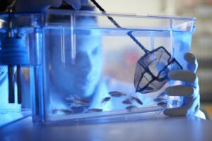
Neck and back pain could be caused by changes in gene activity that trigger the breakdown of the spine’s natural shock absorbers, according to researchers in the U.K. Findings from a study in zebrafish, titled “Targeted modulation of phosphate and lipid metabolism reduces ligament mineralization in col9a1b deficient zebrafish,” and published in Communications Biology, suggest that changes in gene activity can lead to a build-up of minerals in the spine—similar to unwanted bone forming in the wrong place—causing it to harden.
Experts say the findings point to potential future drug targets to treat back pain and suggest zebrafish could be a valuable tool for testing them.
Back pain affects most people at some point in their lives. One of the main underlying causes is the gradual breakdown of spinal discs (which cushion the bones of the spine), known as intervertebral disc degeneration (IVDD).
Despite how common and costly IVDD is, there are currently no drugs that can stop or reverse the condition. Surgery remains the only long-term option.
Genetics are known to play a role in the development of IVDD. A gene connected to a protein called collagen IX, which helps hold the disc’s structural fibers together, has been repeatedly linked to early-onset disc problems.
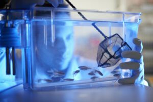
Scientists from the Universities of Edinburgh and Bristol studied zebrafish that were bred to lack a working copy of the gene to better understand how genetic faults could lead to disc disease. As the fish aged, their spines developed problems strikingly similar to human disc disease. The bones of the spine fused together, and the tissue between vertebrae became abnormally hardened with mineral deposits.
The team found that this hardening was preceded by a breakdown in a supportive scaffold layer in the developing spine, well before any mineral began to build up.
Researchers looked at which genes were switched on or off in the fish. They uncovered disruptions to how the body handles fat and to a growth-control pathway called mTOR, alongside changes in phosphate handling and vitamin A signaling, all processes linked to mineral buildup.

The team was also able to demonstrate ways to reduce the damage. The bone-protecting drug bisphosphonate, which is already used for osteoporosis, blocked the mineral buildup. Simply restricting the fish’s food intake, or using drugs that dampen fat metabolism, also reduced spinal fusions.
The findings point to phosphate handling and fat metabolism as promising targets for future drugs, according to the research team.
“For decades, surgery has been the only real answer for disc disease. By understanding the biology that drives the spine to harden, our zebrafish studies point to several ways of slowing it down, including a drug already used safely in patients,” said Erika Kague, PhD, study lead from the University of Edinburgh’s Institute of Genetics and Cancer. “There’s more work to do, but for a condition that’s affected people for generations without a treatment in sight, this is super exciting.”
“For the 9.5 million people across the U.K. living with back pain, this research brings fresh hope that potential new therapeutic approaches are on the horizon,” added Caroline Aylott, PhD, head of research delivery at Arthritis UK. “We are proud to fund research that is unlocking the science behind the processes leading to spinal disc degeneration. Back pain is one of the U.K.’s most common conditions that has blighted millions over generations.
“Dr. Erika Kague and her team at the University of Edinburgh have uncovered important genetic evidence that could pave the way for new treatments, bringing us one step closer to a future where fewer people have to live with the daily pain and challenges that back pain can bring.”
The post Genetic Findings Provide Insights Into Leading Cause of Back Pain appeared first on GEN – Genetic Engineering and Biotechnology News.


The accelerated clearance follows two earlier rejections and the support of an advisory panel that disputed the arguments of FDA scientists.
About 15 to 20 million people globally are impacted by spinal cord injuries, which can impair movement, limit their independence, and disrupt important bodily functions. For example, damage to the spinal cord that occurs at the neck disrupts signals that control the diaphragm, the main muscle used in breathing. The body does not naturally rebuild lost neural connections and there are no approved therapies that can regenerate the neurons and connections affected by a spinal cord injury. But that could change thanks to new research from scientists at Gladstone Institutes.
Full details of the work, which was done in rats, are published in Science Translational Medicine in a new paper “Human spinal interneurons repair the injured rat spinal cord through synaptic integration.” It shows that human stem cell-derived spinal interconnected neurons or interneurons—critical cells for breathing and movement—can survive following transplantation in injured rats, form connections with the receiving animals’ neural circuits, and improve breathing-related motor function. As Lana Zholudeva, PhD, a Gladstone investigator and the paper’s first author, puts it, “this study demonstrates that a specific type of human spinal interneuron can be engineered from stem cells and transplanted into an injured spinal cord” in such a way that “the cells not only survive, but form new pathways to repair damaged networks.”
For the study, the scientists focused on a subtype of the interneurons called V2a interneurons. These are relay cells that play a role in controlling movement. Previous research by Zholudeva’s team and others have shown that these cells are implicated in recovery after traumatic spinal cord injury, including in the neural circuits involved in breathing and walking.
Using human induced pluripotent stem cells, Zholudeva and her team generated transplantable human V2a interneurons that were optimized for repairing injured spinal circuits. Specifically, “we engineered human V2a-enriched SpINs from an optogenetic channelrhodopsin-2 (ChR2) expressing the human induced pluripotent stem cell line,” they wrote in the paper. Getting the process right took some doing, according to Deepak Srivastava, MD, Gladstone president and senior author of the study “it took about a year and a half of trial and error to get the recipe right to make this particular neuron out of stem cells, but it really paid off.” They also ensured that cells could be frozen in vials and later thawed for use, making it possible to use them in human clinical trials down the road.
Next, the scientists transplanted the interneurons into adult rats one week after they sustained injuries to their cervical spinal cords. Two months post transplantation, the scientists found that the new cells not only survived the hostile environment of the injury site but also formed connections with nearby cells. Furthermore, when the scientists activated the transplant site, they observed increased activity in the diaphragm. They also activated the rats’ own brainstem neurons and found that the transplanted cells switched on in response.
The scientists also tested the rats’ breathing under different conditions. Under normal conditions, the difference in the animals’ breathing was less noticeable. But in a low oxygen or high carbon dioxide environment, most of the injured, untreated controls showed signs of respiratory failure. In contrast, most of the rats that received the new V2a interneurons passed the challenges without difficulty. “The transplanted cells seem to be providing that additional capacity,” Zholudeva said.
One component of the study involved looking at why some transplants worked better than others. The scientists identified a specific subset of transplanted V2a interneurons that seemed especially likely to connect with the host animal’s breathing circuit. They plan to follow up on the finding as part of their next steps. Further down the road, they plan to test the potential therapy in larger animals. And they will evaluate whether it is as effective in the injured spinal cord months or years after injury, not just in the immediate aftermath.
The team also hopes to test the treatment in other neural circuits. Specifically, they are considering circuits that control arm and hand function, something that people with cervical spinal cord injuries often identify as their highest priority for recovery. “We’ve shown a proof of principle that this can work, that you can engineer a defined cell type, transplant it, and have it actually repair a specific circuit,” Zholudeva said. “Now we have to make it work more consistently, in more circuits, and eventually in people.”
The post Engineered Human Interneuron Transplants Repair Respiratory Circuits in Injured Rats appeared first on GEN – Genetic Engineering and Biotechnology News.


Illinois’ financial crisis could bring the state to a halt
The final 6 ‘Game of Thrones’ episodes might feel like a full season
New Season 8 Walking Dead trailer flashes forward in time
Mod turns ‘Counter-Strike’ into a ‘Tekken’ clone with fighting chickens
Meet Superman’s grandfather in new trailer for Krypton
Disney’s live-action Aladdin finally finds its stars

Genetic Findings Provide Insights Into Leading Cause of Back Pain

Replimune rebounds to win FDA approval of melanoma drug
Engineered Human Interneuron Transplants Repair Respiratory Circuits in Injured Rats
Third time’s the charm for Replimune as melanoma drug earns FDA greenlight

Tau and Alzheimer’s Disease

STAT+: FDA approves Replimune melanoma drug previously rejected twice
Illinois’ financial crisis could bring the state to a halt
The final 6 ‘Game of Thrones’ episodes might feel like a full season
New Season 8 Walking Dead trailer flashes forward in time
Mod turns ‘Counter-Strike’ into a ‘Tekken’ clone with fighting chickens
Meet Superman’s grandfather in new trailer for Krypton
Disney’s live-action Aladdin finally finds its stars
-
Fierce Biotech4 months ago
Scientists turn pig semen extract into eye drops that kill cancer in mice
-
 Uncategorized9 years ago
These ’90s fashion trends are making a comeback in 2017
-
Endpoints News5 months ago
Novartis to pay $2B upfront to take next-gen PI3Kα inhibitor from Synnovation
-

 Uncategorized5 months ago
Novartis buys Synnovation’s PI3Kα inhibitors for $3 billion
-
Uncategorized9 years ago
According to Dior Couture, this taboo fashion accessory is back
-
Uncategorized9 years ago
Steph Curry finally got the contract he deserves from the Warriors
-
Uncategorized9 years ago
Phillies’ Aaron Altherr makes mind-boggling barehanded play
-
Uncategorized9 years ago
Uber and Lyft are finally available in all of New York State