Uncategorized
6 times when drug development got personal
Uncategorized
STAT+: Human Cell Atlas leader’s tie to 10x Genomics raises conflict-of-interest questions
A decade since its founding, the International Human Cell Atlas Consortium is hosting a high-profile meeting in Boston this week, with panels featuring more than two dozen prominent academics and biotech industry leaders, including Genentech’s Aviv Regev, David Altshuler of Vertex Pharmaceuticals, and Eric Lander from the Broad Institute. The event, which is expected to draw hundreds of scientists from across the globe, comes at an inflection point in the HCA’s ambitious aim to build a comprehensive reference map of all the different types of cells that make up a human body.
Later this year, the HCA expects to deliver a first draft — the completion of single-cell atlases across all of the major organs and tissues — that promise to boost researchers’ understanding of how the body works. So far the HCA has focused on building a reference of healthy cells and the genes they express. In its next phase, it plans to tackle disease, and that means amassing knowledge about where particular cells are located, who their neighbors are, and who they’re communicating with — a rapidly emerging field known as spatial biology.
It’s embarking on this expansion when the field is awash in new technology options from companies like Vizgen, Bruker, Illumina, Takara Bio, Bio-Techne, and 10x Genomics, a Bay Area company whose single-cell RNA sequencing technology was the workhorse of the HCA’s first phase. Scientists who want to join this effort will be faced with decisions about which commercially available solutions to use.
Cancer drugs can shrink fast-growing tumors. But sometimes a few tumor cells survive. These “persister” cells seed new tumors, forcing cancer patients into arduous cycles of testing and treatment. The problem is that persister cells are rare—as few as one in a thousand tumor cells—and they’re genetically identical to the tumor, which makes them hard to find. Plus, their tenacity can be temporary, and by the time a scientist can get them in a petri dish, the qualities that helped them survive may have faded.
To figure out how to beat them, researchers at the University of California, San Francisco (UCSF), built a robotic system that treats thousands of mini tumors at once in the laboratory. Their resulting ResMap platform lets scientists systematically identify, track, and treat surviving cells. The platform revealed shared features among persister cells that could help explain why cancer comes back—features that could be exploited by future drug therapies to beat them. “A few years ago, people were still asking whether persister cells were real,” said Xiaoxiao “Vany” Sun, PhD, an assistant researcher in the UCSF Department of Pharmaceutical Chemistry. “Now we can find them and test ideas for how to eliminate them.”
Sun is first author of the team’s published paper in Science Advances, titled “ResMap: A community resource for systematic mapping of therapy-persistent residual cancer cell dependencies across contexts,” stating, “ResMap establishes a foundation for coordinated community efforts to accelerate rational persister-directed combination strategies toward the clinic.”
Residual disease following targeted therapy remains a key challenge to achieving lasting responses in oncogene-driven cancers, the authors stated. Drug-tolerant persister cells, which the team describes as “subpopulations that survive initial therapy without stable genetic resistance,” can contribute to residual disease and seed tumor relapse. “Targeting drug-tolerant persister cells has emerged as an essential complement to oncogene-directed therapy, yet the field has lacked a unified framework to evaluate and prioritize candidate targets,” they wrote. “Understanding and targeting these cells have emerged as a promising strategy for achieving lasting therapeutic outcomes.”
Cancer cell persistence was first described in 2010, the authors explained, and studies have linked persister survival to different biological processes and resulted in “an expanding list” of candidate therapeutic targets. However, they noted, “… despite over a decade of research, no persister-directed therapy has reached clinical approval.”
For their reported study, the team gathered 94 drug candidates that other laboratories had flagged as potential persister therapies. They wanted to test each drug at different doses, on persisters from two types of lung cancer that had been treated with standard therapies. “As a testbed, we selected four lung cancer models: two with EGFR inhibitor osimertinib (EGFRi)–treated EGFRmut cell lines (PC9 and MGH134) and two with KRAS inhibitor sotorasib (KRASi)–treated KRASG12C cell lines (LU65 and MGH1138-1),” they wrote in summary. Each model was screened under normal oxygen and hypoxic conditions.
It would require 10,000 painstaking, week-long experiments—so they built a robotic platform to eliminate the labor and inconsistency of doing it by hand.
Thousands of miniature tumors sat in stacks of 384-well plates inside controlled incubators. A robotic arm, like those used in pharmaceutical drug screening, moved the plates between experimental stations. One station used sound waves to deposit tiny, precise doses of drug onto each tumor (first, a lung cancer therapy; then, an experimental persister therapy). Other stations stained the tumors with antibodies and took microscopic images of each tumor or group of persisters.
The overall ResMap platform incorporated multiple components, the team explained. “… we developed the ResMap platform incorporating four integrated components: an automated high-throughput workflow, machine learning-based normalization, a persistence-specific metric, and a validated framework.”
Their results showed that of the tested drugs, nine consistently weakened persister cells. The findings suggest that persister cells may share common vulnerabilities, even if they had emerged under different treatment conditions. “Initial screening identified 12 targets with conserved anti-persister activity across genotypes and oxygen environments; follow-up validation reproduced nine of these targets and revealed variable degrees of persister specificity relative to general cytotoxicity.” The investigators suggested that, “Collectively, these findings suggest that although persister biology involves multiple adaptive programs, targeting individual, well-chosen survival pathways may be sufficient to meaningfully reduce residual disease burden.”
Steve Altschuler, PhD, professor of pharmaceutical chemistry at UCSF and co-senior author of the paper, said, “We expected each tumor to behave as its own special case. Instead, we found patterns that held up across many different samples, suggesting there may be underlying rules that can help predict which therapies are most likely to work.”
The team plans to expand the platform to include more tumor types and treatment conditions. They hope the resulting dataset will be a resource to help researchers eliminate persister cells before they can give rise to drug-resistant disease. “ResMap provides a community resource for coordinated validation efforts and rational combination design aimed at minimizing residual disease following anticancer therapy,” they stated.
The post Therapy-Resistant Residual Cancer Cell Dependencies Mapped appeared first on GEN – Genetic Engineering and Biotechnology News.

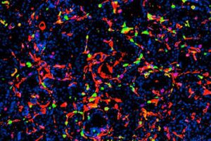
Ultrasmall fluorescent core‑shell silica nanoparticles—best known for their roles in medical imaging applications—are now showing surprising therapeutic muscle. Originally engineered as inert carriers for imaging agents, these particles, called Cornell Prime dots (C’ dots), have steadily expanded their résumé. In a new preclinical study, researchers at Weill Cornell Medicine report that these engineered silica nanoparticles can directly kill prostate tumor cells while reawakening antitumor immunity, offering a potential new edge in a disease where immunotherapy has historically struggled.
Prostate cancer remains one of the most immunologically “cold” solid tumors, with myeloid‑driven immune suppression, metabolic bottlenecks, and stromal remodeling that blunt the effects of checkpoint blockade. The new work suggests that C’ dots—when targeted to prostate‑specific membrane antigen (PSMA)—can break through these layers of resistance by triggering ferroptosis, remodeling the tumor microenvironment, and priming tumors for combination immunotherapy.
“We’re very encouraged by these results; a treatment that directly induces tumor‑cell death while transforming the immune microenvironment, as this does, would represent a new clinical paradigm,” said senior author Michelle Bradbury, MD, PhD, the endowed professor of imaging research in radiology and director of the Molecular Imaging Innovations Institute at Weill Cornell Medicine and a neuroradiologist at NewYork-Presbyterian/Weill Cornell Medical Center.
The study, published in Cancer Research and titled “Reprogramming of TLR–Ferroptosis Signaling and Immunometabolic Pathways Overcomes Myeloid Suppression to Improve Checkpoint Blockade in Prostate Cancer,” shows that the silica particles accumulate in prostate tumors and push cancer cells toward ferroptosis, a form of iron‑dependent cell death driven by runaway lipid peroxidation. Although the particles were originally designed for imaging, the team found that they often pick up positively charged iron ions in the bloodstream and shuttle them into tumor cells—effectively turning the particles into catalytic seeds for oxidative collapse.
At the same time, the nanoparticles reshape the immune landscape. T cells, macrophages, and other immune populations shift from inert or suppressive states into robust antitumor activity, converting cold tumors into hot ones. “One of the most intriguing aspects of this work is the convergence of direct tumor cell killing with broad immune remodeling,” said co‑author Jedd Wolchok, MD, PhD, the Meyer director of the Sandra and Edward Meyer Cancer Center, professor of medicine at Weill Cornell Medicine, director of the Parker Institute for Cancer Immunotherapy at Weill Cornell Medicine Meyer Cancer Center, and an oncologist at NewYork-Presbyterian/Weill Cornell Medical Center.
The therapeutic impact was most striking in survival experiments. C’ dots alone modestly extended survival in aggressive mouse models, as did checkpoint blockade alone. But the combination produced complete or near‑complete remissions in 40% of mice. Adding CSF‑1R blockade increased complete remissions to 50%.
The researchers’ next steps include continuing to explore these ultrasmall core-shell silica particles, setting the stage for the platform’s translational potential.
“By creating conditions that support a more effective antitumor immune response, these particles may help unlock the full potential of immunotherapy in prostate cancer, where durable responses have historically been difficult to achieve,” added Wolchok.
The post Silica Nanoparticles Induce Ferroptosis, Reprogram Immunity in Prostate Cancer Models appeared first on GEN – Genetic Engineering and Biotechnology News.


Illinois’ financial crisis could bring the state to a halt
The final 6 ‘Game of Thrones’ episodes might feel like a full season
New Season 8 Walking Dead trailer flashes forward in time
Mod turns ‘Counter-Strike’ into a ‘Tekken’ clone with fighting chickens
Meet Superman’s grandfather in new trailer for Krypton
Disney’s live-action Aladdin finally finds its stars

STAT+: Human Cell Atlas leader’s tie to 10x Genomics raises conflict-of-interest questions
6 times when drug development got personal
Therapy-Resistant Residual Cancer Cell Dependencies Mapped

Silica Nanoparticles Induce Ferroptosis, Reprogram Immunity in Prostate Cancer Models

When Process Design Fails: 5 Common Planning Gaps That Create Downstream Purification Bottlenecks
mRNA Flu Vaccine Shows Stronger, Longer-Lasting Immune Response
Illinois’ financial crisis could bring the state to a halt
The final 6 ‘Game of Thrones’ episodes might feel like a full season
New Season 8 Walking Dead trailer flashes forward in time
Mod turns ‘Counter-Strike’ into a ‘Tekken’ clone with fighting chickens
Meet Superman’s grandfather in new trailer for Krypton
Disney’s live-action Aladdin finally finds its stars
-
 Uncategorized9 years ago
These ’90s fashion trends are making a comeback in 2017
-
Uncategorized9 years ago
According to Dior Couture, this taboo fashion accessory is back
-
Endpoints News3 months ago
Novartis to pay $2B upfront to take next-gen PI3Kα inhibitor from Synnovation
-
Uncategorized9 years ago
Phillies’ Aaron Altherr makes mind-boggling barehanded play
-
Uncategorized9 years ago
Uber and Lyft are finally available in all of New York State
-
Contributors9 years ago
The final 6 ‘Game of Thrones’ episodes might feel like a full season
-
Uncategorized9 years ago
Steph Curry finally got the contract he deserves from the Warriors
-
Uncategorized9 years ago
The old and New Edition cast comes together to perform