Uncategorized
Target for Aggressive Prostate Cancer Prevention Identified in Mice
Researchers at Columbia University Irving Medical Center have identified a gene that drives the development of neuroendocrine prostate cancer (NEPC), an aggressive form of the disease. Their study showed that genetic or pharmacological inhibition of Sirtuin 1 prevents the growth of NEPC tumors in mice, laying the groundwork for future clinical studies aimed at developing new treatments for NEPC in humans.
Research lead Cory Abate-Shen, PhD, a professor at Columbia University Vagelos College of Physicians and Surgeons, is co-senior author of the researchers’ published paper in Journal of Experimental Medicine, titled “A forward genetic screen identifies Sirtuin 1 as a driver of neuroendocrine prostate cancer,” in which the team noted, “We demonstrate that expression of Sirt1 promotes NEPC while its silencing or pharmacological inhibition suppresses the NEPC phenotype.”
Prostate cancer is the most common cancer in men, and one in every six men will be affected by prostate cancer in their lifetime, the authors explained. Prostate cancer treatments have been focused on approaches to dampening androgen receptor (AR) signalling, and for men with recurrent or advanced prostate cancer the current standard of care is androgen deprivation therapy (ADT). However, it is well documented that ADT will eventually fail, leading to tumor recurrence and development of the ADT-insensitive aggressive prostate cancer variant, NEPC. “… while ADT initially leads to tumor regression, eventually tumors recur as castration-resistant prostate cancer (CRPC), so called because of its continued reliance on AR even in the absence of androgens,” the team continued.
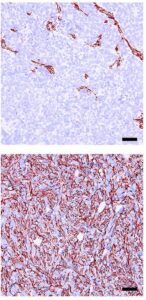
![Positively stained NEPC markers (top) are lost when Sirt1 is silenced (bottom). [© 2026 Nunes de Almeida et al. Originally published in <em>Journal of Experimental Medicine</em>. https://doi.org/10.1084/jem.20241484]](https://www.genengnews.com/wp-content/uploads/2026/05/Low-Res_Nunes_de_Almeida_et_al_2-146x300.jpg)
The process through which ADT-responsive tumors transition towards NEPC tumors—a phenomenon known as lineage plasticity—remains unknown. “Elucidating the mechanisms governing this process may improve treatment by overcoming plasticity-associated drug resistance,” Abate-Shen added.
For their newly reported study the research team performed a Sleeping Beauty (SB) forwards genetic mutagenesis screen in mice looking for mutations that recurred across multiple independent prostate cancer tumors. They identified 75 candidate NEPC-promoting genes, the most promising of which was Sirtuin 1 (Sirt1). Sirt1 encodes an enzyme with a broad range of functions, including control of gene expression and metabolism.
The group first looked to a human prostate cancer cell line to characterize the role of Sirt1. In these cells, the induction of NEPC produced an increase in the expression of genes predicted to be activated by SIRT1 and a corresponding decrease in those predicted to be downregulated by this protein. Confirming these results, the group found that activation of Sirt1 in cells with low SIRT1 expression levels led to a robust increase in key NEPC markers.
Recapitulating their cell line data, the team found that silencing of Sirt1 profoundly reduced tumor growth in mice with NEPC, indicating that Sirt1 is indeed a promising target for NEPC treatment. They also treated the tumors with the FDA-approved SIRT1-inhibitor, Selisistat, which was originally developed for treatment of Huntington’s disease. Encouragingly, the researchers saw that Selisistat administration significantly reversed the NEPC phenotype. “Functional studies in human prostate cancer cell models and mouse organoid models using gain- and loss-of- function approaches in vitro and in vivo, as well as pharmacological inhibition, demonstrated that one of the top-ranked candidates, Sirt1, promotes NEPC,” they wrote in summary.
![Tumors surgically removed from Sirt1-silenced mice (bottom) are significantly smaller than tumors removed from mice with wild-type Sirt1 expression. [© 2026 Nunes de Almeida et al. Originally published in <em>Journal of Experimental Medicine</em>. https://doi.org/10.1084/jem.20241484]](https://www.genengnews.com/wp-content/uploads/2026/05/Low-Res_Nunes_de_Almeida_et_al_1-300x275.jpg)
“Our findings demonstrate that SIRT1 plays a pivotal role in promoting NEPC, revealing a context-dependent function that extends beyond general tumor growth to the regulation of lineage plasticity and neuroendocrine differentiation,” says Abate-Shen, adding that “this highlights SIRT1 as an attractive and clinically actionable target for lethal prostate cancer that warrants further investigation in future clinical studies.”
In their paper the team concluded, “Overall, our study establishes a generalizable computational and experimental framework that integrates SB mutagenesis with phenotypic, genomic, and transcriptomic analyses to identify novel cancer drivers … Importantly, our data suggest that targeting SIRT1 may suppress or reverse progression toward NEPC.”
The post Target for Aggressive Prostate Cancer Prevention Identified in Mice appeared first on GEN – Genetic Engineering and Biotechnology News.
Uncategorized
STAT+: At hospital finance conference, a call to end the friction that’s keeping costs high
NATIONAL HARBOR, Md. — At this week’s annual meeting of hospital finance leaders, the exhibit hall was packed with dozens of billing and collections companies. Armed with candy, tote bags, and pens, they smiled at passersby, eager to explain why their tactics would extract the most money from health insurers.
The sheer number of “revenue cycle” vendors who attended the Healthcare Financial Management Association’s annual conference in Maryland — outnumbering even the hospital attendees, according to a list shared by an organizer — was a visible reminder of the enormous industry built around just paying medical bills.
The U.S. health care industry spends roughly $200 billion annually on financial transactions: claims processing, payment, collections, and prior authorization. And yet the proliferation of billing vendors seemed to clash with the main theme of HFMA’s conference, affordability, spotlighting the need to simplify the billing process so that health care is less costly and more accessible for patients.
Strong science, lower costs and growing capital networks are putting Spain and Portugal on the biotech investment map, even as structural bottlenecks persist, according to two investors.

You know when you are at the eye doctor getting an updated prescription, and suddenly the world snaps into sharper focus? Physicists at the University of California (UC), Berkeley, have now done something similar for electron microscopy. By introducing phase contrast into a cryo‑electron microscope, they have delivered dramatically sharper images of some of biology’s smallest and most elusive proteins.
The advance comes from a new laser phase plate (LPP), described in the paper “Laser phase plate improves structure determination of small proteins by cryo‑EM,” which was published recently in Science. Led by physicist Holger Mueller, PhD, of UC Berkeley and Lawrence Berkeley National Laboratory, the team demonstrated that a laser‑driven phase plate can overcome one of cryo‑EM’s most persistent limitations: poor contrast for small proteins.

Cryo‑EM has transformed structural biology over the past decade, earning a Nobel Prize in 2017 for enabling high‑resolution structures without crystallization. But despite its impact, the technique still struggles with proteins below ~70 kilodaltons—a size range that includes about 90% of the human proteome. “Because of signal-to-noise limitations, the majority of human and animal proteins are too small to be analyzed by these methods [cryo-EM and cryoelectron tomography]. The increase in signal-to-noise ratio provided by this laser phase plate is expected to overcome these important limitations.”
The new LPP begins to address that problem. The LPP uses an intense, continuous‑wave laser to shift the phase of the electron beam itself. This produces true phase contrast without dimming or destabilizing the beam. Mueller described the laser focus as “75 kilowatts focused to a few microns… That’s more powerful than what you use for welding. It has more power than a military laser. It builds up the brightest continuous laser focus ever.”
Installed in a custom Thermo Fisher Titan Krios, the LPP immediately improved the clarity and resolvability of small proteins, including hemoglobin, which sits at the lower limit of what today’s cryo‑EM instruments can handle. As the authors wrote in the abstract: “Here, we show that the laser phase plate (LPP)… enhances the resolution in single-particle reconstruction of small proteins by improving specimen-motion correction, recovery of information from the early frames, as well as particle visualization, 3D classification, and alignment.”

These improvements were achieved using standard defocus ranges and reconstruction workflows. “For the most challenging cases—small particles, bad specimens—the laser produces a very considerable advantage,” Mueller said.
The impact extends beyond single‑particle analysis. Cryo‑electron tomography (cryo‑ET), which assembles multiple angular views of a molecule or protein into a three-dimensional image, stands to benefit even more. “With cryo-ET, we’re looking at small, very complicated cellular material that’s incredibly crowded inside the cell,” said Bridget Carragher, PhD, founding technical director of imaging at Biohub. “It’s like a forest of trees, and you’re trying to find one leaf on one tree in there. Cryo-ET needs a dramatic step forward in contrast, so we can start to see what’s going on inside the cell. That’s what the laser phase plate promises to give us.”
Biohub is developing a dual‑laser version of the system, designed to reduce component wear and minimize aberrations. Meanwhile, Mueller’s team is pushing toward imaging proteins as small as 17 kilodaltons, a threshold that would open access to vast regions of the human proteome previously invisible to cryo‑EM.
“This technology is a step function change for biology,” said Stephani Otte, PhD, Biohub’s vice president of imaging science. “What was once invisible will become visible—and that changes everything about how we understand disease.”
“The bottom line is, if you have a large protein and a really good sample—a fresh one or one frozen without bubbles, for example—you may not need the phase plate to get a single, high-quality image. But for a small protein and a bad sample, laser-on is best,” Mueller said. “This could fill an enormous gap in our knowledge of protein structures that can’t be crystallized or are too small for today’s cryo-EM. And it will be revolutionary for cryo-ET.”
The post Laser‑Driven Phase Contrast Enhances Cryo‑EM Resolution of Small Proteins appeared first on GEN – Genetic Engineering and Biotechnology News.


Illinois’ financial crisis could bring the state to a halt
The final 6 ‘Game of Thrones’ episodes might feel like a full season
New Season 8 Walking Dead trailer flashes forward in time
Mod turns ‘Counter-Strike’ into a ‘Tekken’ clone with fighting chickens
Meet Superman’s grandfather in new trailer for Krypton
Disney’s live-action Aladdin finally finds its stars

STAT+: At hospital finance conference, a call to end the friction that’s keeping costs high
Beyond sunshine: Iberia’s biotech moment has arrived with developing capital networks

Laser‑Driven Phase Contrast Enhances Cryo‑EM Resolution of Small Proteins

STAT+: Updated: Tracking RFK Jr.’s promises to remake health in America

An obesity drug deep-dive, and peptides move mainstream

RFK Jr. claims his calendar is publicly available. We’ve been trying to get it for a year
Illinois’ financial crisis could bring the state to a halt
The final 6 ‘Game of Thrones’ episodes might feel like a full season
New Season 8 Walking Dead trailer flashes forward in time
Mod turns ‘Counter-Strike’ into a ‘Tekken’ clone with fighting chickens
Meet Superman’s grandfather in new trailer for Krypton
Disney’s live-action Aladdin finally finds its stars
-
 Uncategorized9 years ago
These ’90s fashion trends are making a comeback in 2017
-
Uncategorized9 years ago
According to Dior Couture, this taboo fashion accessory is back
-
Endpoints News3 months ago
Novartis to pay $2B upfront to take next-gen PI3Kα inhibitor from Synnovation
-
Uncategorized9 years ago
Phillies’ Aaron Altherr makes mind-boggling barehanded play
-
Uncategorized9 years ago
Uber and Lyft are finally available in all of New York State
-
Contributors9 years ago
The final 6 ‘Game of Thrones’ episodes might feel like a full season
-
Uncategorized9 years ago
Steph Curry finally got the contract he deserves from the Warriors
-
Uncategorized9 years ago
The old and New Edition cast comes together to perform