Uncategorized
Synthetic Biology and Tissue Engineering Grow Liver Tissue In‑Body
Damage to the liver in patients developing end-stage liver disease has become too severe for the organ’s normally extraordinary regenerative capacity to repair or compensate for that damage. Once this point of no return has been reached the only option is an organ transplant. However, donor livers are in high demand and very limited supply.
Ambitious efforts are on the way that eventually could enable the engineering of entire implantable liver organs. However, the maximum size of laboratory-engineered liver constructs remains limited and cannot yet provide therapeutic benefits for patients. A research team at the Wyss Institute at Harvard University, Boston University, and MIT has now approached this important problem from a different angle.
“We asked if it would be possible to first implant a small-scale liver construct and then drive it to expand in the body following its engraftment,” said Christopher Chen, MD, PhD, a Wyss Institute core faculty member and the William Fairfield Warren Distinguished professor of biomedical engineering and director of the Biological Design Center at Boston University. “A sufficiently grown, functional ‘satellite liver’ could immediately relieve the metabolic burden in a damaged liver and help bridge the time until a transplant becomes available.”
Chen co-led the research together with associate faculty member Sangeeta Bhatia, MD, PhD, who is the John J. and Dorothy Wilson Professor of Health Sciences and Technology and of Electrical Engineering and Computer Science at the Koch Institute for Integrative Cancer Research at MIT, and a Howard Hughes Medical Institute investigator. Chen is also a leader of the Wyss Institute’s 3D Organ Engineering Initiative, and team lead of the recently awarded ARPA-H PRINT-supported ImPLANT project, which focuses on whole organ liver engineering at the Wyss and collaborating institutions.
The project, spearheaded by Amy Stoddard, PhD, (MIT ’25), who developed the approach in her doctoral research and then as a postdoctoral fellow, integrates tissue engineering and synthetic biology tools in a genetic strategy the team has named “bioengineered on-demand outgrowth via synthetic biology triggering,” or BOOST. By specifically rewiring the gene expression of primary liver hepatocytes and supportive fibroblast cells, the scientists were able to effectively switch on a tissue growth program in small, engineered liver constructs after their implantation into mice.
“Using engineered liver tissue as a proof-of-concept application, we integrated synthetic biology and tissue engineering tools to build liver tissues that can be expanded on-demand after implantation in vivo,” the team reported in their published paper in Science Advances, which is titled “Synthetic control of implanted engineered liver tissue growth.” In the paper they concluded “In this study, we define the first steps toward an unconventional approach to cell therapy scale-up: engineering a small construct and then inducing it to grow in situ … “This strategy, which we have named BOOST, could provide several key advantages, including circumventing the need for large quantities of cellular raw materials and bypassing the formidable challenge of generating a rapidly perfusable construct that can survive the engraftment period.”
The authors wrote, “Organ transplant is currently the only curative treatment for patients with end-stage organ failure, yet this therapy is inaccessible to many due to the paucity of organs available for transplant.” And while significant progress has been made in the field of engineering tissue-based cell therapies that could represent alternatives, or bridges to transplant, they acknowledge, “… scaling of these constructs to sizes of therapeutic relevance remains a barrier to clinical translation.”
In order to address current challenges associated with fabrication, Chen and colleagues looked at the problem from different angle, asking whether it would be possible to first implant a small-scale construct and then trigger it to expand in situ, after its engraftment into the host.
To be able to induce growth of an implanted small liver constructs in situ within a recipient’s body the researchers first needed to identify the relevant cues that would allow them to do so. “A key first step toward this method of in situ scale-up would be the successful control of cellular growth within the engineered construct after engraftment,” they wrote. Since liver growth is known to be regulated by soluble growth factors (GFs), Stoddard screened a collection of candidate factors to identify those that, when added to cultured human primary hepatocyte cells (HEPs), had the strongest growth-inducing effects.
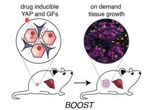
![The genetic “BOOST” strategy integrates tissue engineering and synthetic biology tools to enable on-demand liver growth inside the body. By specifically rewiring the gene expression of primary liver hepatocytes and supportive fibroblast cells, a tissue growth program is switched on in a small, engineered liver construct after its implantation into recipients and upon addition of an inducing agent (shown as a pill). As a result, the hepatocytes in the construct start and continue to proliferate until a desired construct size has been reached and the inducing signal is not provided anymore. In mice, BOOST resulted in robust and healthy liver growth. [Wyss Institute at Harvard University]](https://www.genengnews.com/wp-content/uploads/2026/04/Low-Res_Press-graphics-01-300x225.jpg)
“We ended up with a set of four growth factors, HGF, TGFa, WNT2 and RSPO3, that potently induced sparsely scattered HEPs to grow in the culture dish,” said Stoddard. “But when we tested whether they could do the same in 3D liver tissues consisting of densely packed HEPs and fibroblasts, they turned out to be ineffective. This led us to hypothesize that there must be an additional mechanism at work in human HEPs that inhibits cell proliferation in high-density conditions.”
The team homed in on a protein, YAP, that senses mechanical signals, and which was known to move from cells’ cytosol to their nucleus in low-density conditions to help express genes involved in cell proliferation. However, in high-density conditions when cells are compressed, YAP is degraded in the cytosol, which prevents the activation of those target genes and restricts proliferation.
“Importantly, when we overexpressed a non-degradable version of YAP in HEPs, which also reaches the nucleus in high-density conditions to partake in gene regulation, we successfully overrode this density checkpoint in HEPs,” Stoddard said. “Interestingly, we found that HEPs needed to be stimulated with both YAP and GFs in order to grow in densely packed 3D liver tissues.”
Toward the goal of safely inducing and controlling HEP proliferation in a living organism, and eventually human patients, the researchers deployed synthetic biology tools to locally install control of these signaling pathways in HEPs and fibroblast cells within the engineered 3D liver tissues themselves. “We set out to engineer a synthetic biology toolkit capable of locally modulating growth factor and YAP signaling within engineered liver tissue, enabling on-demand control of proliferation even after implantation,” they noted.
The team engineered fibroblast cell lines that each secreted one of the four GFs, and HEPs that expressed the non-degradable YAP protein. And they made the expression of all proteins doxycycline (DOX)-inducible. They determined in time course experiments that a continuous seven-day treatment with DOX led 3D liver tissue composed of engineered cells to robustly expand in size and cell numbers in the culture dish. On DOX removal the HEPs reverted back to a non-proliferating state.
However, Stoddard noted, “… when we compared the gene expression of single cells in BOOST-engineered, DOX-induced 3D liver tissue to that of cells in non-engineered or BOOST-engineered, non-induced 3D liver tissue, we noticed that the expansion came with a trade-off: high proliferation rates went hand in hand with a less functional HEP state. While we believe this is a natural trade-off seen in a wide variety of biological settings, we hope to be able to address this in the future, recognizing that the liver also has native re-functionalization signals to harness.”
The litmus test for BOOST-engineered growth in 3D liver tissues was to see whether they would similarly expand following their implantation into living mice that were systemically treated with DOX for the same seven-day duration. Experiments showed that the implanted tissue exhibited a striking 500% increase in proliferation with a doubling of the engineered HEPs alone, and was vascularized to accommodate the metabolic demands of the expanded tissue. The tissue implants were also well tolerated by the mice, with no signs of fibrosis due to invading immune cells and fibroblast inflammation, or of tumor growth.
“These results were particularly exciting to us,” said Stoddard. “Prior to our work, injury to the host liver has always been required to trigger hepatocyte engraftment and proliferation. Here we were able to relieve this dependence, and trigger on-demand growth of implanted liver tissue in a completely healthy host.”
In the future, the team will explore the capacity of BOOSTed liver tissue to rescue the host in the setting of liver injury. “Our BOOST strategy lays the foundation for a future when solid organ cell therapies can be controlled non-surgically according to the needs of patients and their physicians,” Bhatia noted. “Beyond treating liver disease, the premise of BOOST could be applied to other engineered tissue therapeutics that are similarly constrained by challenges associated with tissue scale-up, such as engineered heart or pancreatic tissue to address serious diseases.”
In their paper the authors concluded, “… this work serves as an exciting proof-of-concept demonstration that scale-up of tissues via growth could be possible … Together, this work helps lay the foundations for a future of ‘smart’ tissue therapeutics that can be scaled to a patient’s needs and thereby offer treatment for numerous, previously incurable, diseases.”
The post Synthetic Biology and Tissue Engineering Grow Liver Tissue In‑Body appeared first on GEN – Genetic Engineering and Biotechnology News.
Last month, the Andes virus outbreak on a Dutch cruise ship departing from Argentina brought a transmission context for hantavirus, that was previously unprecedented, to the forefront. The Andes virus is the only member of the hantavirus family that is capable of efficient person-to-person spread through close contact with respiratory secretions. Other hantaviruses are typically spread through contact with infected rodents, making the Andes virus a much more significant public health threat.
While at sea, the outbreak spread among passengers and crew, infecting 13 people and killing three. The cruise passengers have since returned to their home countries, 23 in total. Because a person can carry the virus for weeks before showing any symptoms, health agencies are facing a complex challenge of identifying everyone who was exposed. There are currently no vaccines or preventive treatments approved for the virus; this travel-related outbreak brought the need for vaccine development to the forefront.
Researchers at The University of Texas Medical Branch (UTMB) had previously developed and tested two mRNA vaccines against intramuscular Andes virus challenge in golden Syrian hamsters (“1-methylpseudouridine-modified or non-modified mRNA modalities encoding the envelope glycoproteins, Gn and Gc, in a single open reading frame.”)
When tested in the Syrian hamster model, both mRNA vaccines were efficacious in hamsters using a two-dose regimen. Recognizing that a fast-moving international outbreak doesn’t allow time for patients to wait weeks between shots, the team retested the vaccines to determine whether a single dose would be effective.
Now, a new report shares the finding that the vaccine provided full protection against the Andes hantavirus after a single dose.
This work is published in The Lancet in the paper, “Single-dose mRNA vaccines against Andes hantavirus.”
Alexander Bukreyev, PhD, head of the Laboratory of Viral Pathogenesis and Vaccine Development at UTMB, said that the group is working to fast-track these single-dose vaccines into human clinical trials.
The results exceeded expectations. When testing the vaccines in an animal model that mimics human disease, the scientists found that a single shot provided 100% protection against a lethal dose of the virus. Even when the researchers significantly lowered the dosage to a fraction of the original amount, the results remained definitive.
“Every vaccinated animal remained completely healthy and showed no symptoms or weight loss,” said Michelle Meyer, PhD, senior scientist in the Bukreyev Laboratory. “When we looked at the tissues from the vaccinated animals a month after infection, the virus was entirely gone. The vaccines triggered a powerful immune response, creating protective antibodies in as little as 14 days.”
Because the Andes virus can take a relatively long time to make a human severely ill, these fast-acting vaccines could serve a dual purpose, possibly functioning as an emergency tool for people who have already been exposed.
“If given quickly to high-risk contacts during an outbreak, such as the Andes virus situation on the cruise ship, the vaccines could theoretically jump-start their immune systems fast enough to intercept the virus—stopping it from replicating and preventing them from getting sick or spreading it further,” Bukreyev said.
The post Hantavirus One-Shot mRNA Vaccine Fully Protects in Syrian Hamster Model appeared first on GEN – Genetic Engineering and Biotechnology News.
Biotechnology company SonoThera has raised $125 million in an oversubscribed Series B financing round. The financing was led by Vida Ventures, with participation from ARK Invest, CureDuchenne Ventures, Leaps by Bayer, Otsuka Pharmaceutical, SymBiosis, UCB Ventures SA, Vivo Capital, and existing investors ARCH Venture Partners, Alexandria Venture Investments, Duquesne Family Office, Illumina Ventures, Johnson & Johnson Innovation – JJDC, Medical Excellence Capital, RA Capital, and Vertex Ventures HC.
SonoThera will use the funds to advance its lead programs in Duchenne muscular dystrophy (DMD) and autosomal dominant polycystic kidney disease (ADPKD) in the clinic. The funds will also support efforts to expand its pipeline of targeted redosable genetic medicines across multiple organ systems and scale its proprietary platform technologies for safe, targeted therapy delivery.
The company’s platform combines a proprietary ultrasound-mediated delivery technology dubbed RIPPLE , with a payload engineering platform dubbed PORE. The platforms are designed to support the development of DNA and RNA therapeutics, gene editing, and gene silencing approaches. SonoThera is using its tech to develop genetic medicines that it claims will address key limitations of conventional gene therapies including delivery challenges, payload size constraints, immune responses, safety events, and difficulties with redosing.
As Kenneth Greenberd, PhD, SonoThera’s co-founder and CEO, stated “we founded SonoThera to take a fundamentally different approach, with a platform designed to broaden the therapeutic possibilities of the field. We believe our technology has the potential to expand the range of diseases addressable by genetic medicines while enabling more precise, durable, safer, and repeatable therapies for patients.”
SonoThera has already demonstrated the targeted delivery and expression capabilities of its platform across multiple tissues, including skeletal muscle, heart, liver, kidney, adipose, and brain. It has also shown that it can deliver large payloads such as full-length dystrophin for DMD and RNA-based payloads for gene silencing applications in preclinical studies.
The company expects to initiate its first clinical trial in DMD in 2027.
Commenting on the financing, Rajul Jain, MD, managing director at Vida Ventures, said “we believe SonoThera, with its RIPPLE delivery and PORE payload engineering technologies, has the potential to unlock opportunities in diseases with significant unmet need that have been previously inaccessible to other genetic medicine approaches.”
In connection with the financing, Jain and Rakhshita Dhar, MS, vice president & head of Healthcare Venture Investments at Leaps by Bayer, have joined SonoThera’s Board of Directors.
The post SonoThera Raises $125M to Develop Ultrasound-Mediated Genetic Medicines appeared first on GEN – Genetic Engineering and Biotechnology News.
Uncategorized
STAT+: Up and down the ladder: The latest comings and goings
Hired someone new and exciting? Promoted a rising star? Finally solved that hard-to-fill spot? Share the news with us, and we’ll share it with others. That’s right. Send us your changes, and we’ll find a home for them. Don’t be shy. Everyone wants to know who is coming and going.
And here is our regular feature in which we highlight a different person each week. This time around, we note that AstronauTx hired Michelle Mellion as chief medical officer. Previously, she held the same role at PepGen and EveryONE Medicines.
But all work and no play can make for a dull chief medical officer.


Illinois’ financial crisis could bring the state to a halt
The final 6 ‘Game of Thrones’ episodes might feel like a full season
New Season 8 Walking Dead trailer flashes forward in time
Mod turns ‘Counter-Strike’ into a ‘Tekken’ clone with fighting chickens
Meet Superman’s grandfather in new trailer for Krypton
Disney’s live-action Aladdin finally finds its stars
Hantavirus One-Shot mRNA Vaccine Fully Protects in Syrian Hamster Model
SonoThera Raises $125M to Develop Ultrasound-Mediated Genetic Medicines

STAT+: Up and down the ladder: The latest comings and goings
FDA imposes import alert on Indian plant after inspectors flag GMP failings

STAT+: Pharmalittle: We’re reading about a discontinued cancer drug, a Novo security breach, and more
Amgen shores up Tavneos’ FDA defense with Duke data analysis
Illinois’ financial crisis could bring the state to a halt
The final 6 ‘Game of Thrones’ episodes might feel like a full season
New Season 8 Walking Dead trailer flashes forward in time
Mod turns ‘Counter-Strike’ into a ‘Tekken’ clone with fighting chickens
Meet Superman’s grandfather in new trailer for Krypton
Disney’s live-action Aladdin finally finds its stars
-
 Uncategorized9 years ago
These ’90s fashion trends are making a comeback in 2017
-
Uncategorized9 years ago
According to Dior Couture, this taboo fashion accessory is back
-
Endpoints News3 months ago
Novartis to pay $2B upfront to take next-gen PI3Kα inhibitor from Synnovation
-
Uncategorized9 years ago
Phillies’ Aaron Altherr makes mind-boggling barehanded play
-
Uncategorized9 years ago
Uber and Lyft are finally available in all of New York State
-
Contributors9 years ago
The final 6 ‘Game of Thrones’ episodes might feel like a full season
-
Uncategorized9 years ago
Steph Curry finally got the contract he deserves from the Warriors
-
Uncategorized9 years ago
The old and New Edition cast comes together to perform