

Uncategorized
Single-Cell Atlas of Maternal–Fetal Interface Sheds Light on Pregnancy Complications

The biological connection between a pregnant woman and her developing baby—the human maternal–fetal interface—is a specialized, transient organ composed of uterine cells from the mother and fetal cells that acts as a barrier, supports fetal growth, and maintains the mother’s health. The cellular complexity of the maternal-fetal interface has limited scientists’ ability to study how healthy pregnancies develop and why complications arise. The underlying cellular, molecular, and spatial programs of the interface—which forms about a week after fertilization and lasts until birth—has remain incompletely defined.
Now, the human maternal–fetal interface has been mapped in unprecedented detail by scientists at the University of California, San Francisco (UCSF), revealing new cell types and providing insights into conditions such as preeclampsia, preterm birth, and miscarriage.
“By examining this tissue cell by cell across pregnancy, we can begin to understand both normal development and what may go wrong,” said Susan J. Fisher, PhD, professor of obstetrics, gynecology, and reproductive sciences at UCSF.
The team generated a comprehensive atlas of the human maternal–fetal interface across normal pregnancies, from early gestation to term. The researchers did this by “integrating large-scale paired single-nucleus transcriptomic and chromatin accessibility profiling with submicrometer-resolution spatial transcriptomics and CODEX multiplex protein imaging.”
Using these tools, the researchers analyzed about 200,000 individual cells and compared them with nearly one million cells in their original positions within the uterine and placental tissue. This enabled them to identify different cell types, track how they develop, and see how they are linked to pregnancy complications.
“This work gives us a much clearer picture of this critical region than ever before,” said Jingjing Li, PhD, associate professor in UCSF’s Department of Neurology and the Eli and Edythe Broad Center of Regenerative Medicine and Stem Cell Research.
This work is published in Nature in the paper, “Single-Cell Spatiotemporal Dissection of the Human Maternal–Fetal Interface.”
The atlas revealed a previously unknown maternal cell type located where fetal placental cells first enter the uterus. These cells appear to regulate how deeply placental cells invade uterine tissue, a process that is essential for establishing blood flow to the fetus. The researchers found that these cells carry a cannabinoid receptor, and exposure to cannabinoid molecules caused them to further restrict placental cell invasion.
“Population studies have linked cannabis use during pregnancy to poorer outcomes,” said Cheng Wang, PhD, a postdoctoral fellow at UCSF. “This cell type may help explain the biological basis of that association.”
To understand how complications arise, the team integrated genetic data from more than 10,000 patients. They mapped genetic risk signals for conditions including preterm birth, preeclampsia, and miscarriage onto regulatory regions of DNA that control gene activity. This approach allowed the researchers to identify the specific cell types and states most strongly associated with each condition.
The team then focused on preeclampsia, a potentially life-threatening disorder marked by sudden high blood pressure. They found that the most affected cell types are involved in remodeling the mother’s uterine blood vessels, a process required to supply adequate blood to the placenta. The findings suggest that preeclampsia may result from disrupted communication between maternal and fetal cells that normally coordinate this process.
Having established a detailed map of healthy pregnancies, the researchers plan to study complicated pregnancies to identify potential targets for treatment.
The post Single-Cell Atlas of Maternal–Fetal Interface Sheds Light on Pregnancy Complications appeared first on GEN – Genetic Engineering and Biotechnology News.

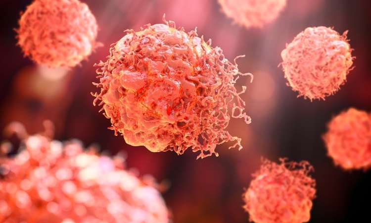

Hosted by Drug Discovery World and supported by Sartorius and BioIVT, this webinar will explore the opportunities and challenges that exist within cancer research drug discovery and development.
You will hear from Dr Sudha Rao, Chief Scientific Officer of Kazia Therapeutics, Karol Budzik, PhD, Business Development Associate at Vyriad Therapeutics and Lars van der Veen, Chief Scientific Officer at iOnctura.
Presentations will cover how cancer treatments have shifted towards reprogramming the biology driving tumour growth, immune escape and treatment resistance, the trajectory that in vivo CAR-T treatments are taking, and how challenging tumours burdened by stroma and immune-mediated resistance can be tackled.
Q&A with the speakers follows the presentations.
The post New webinar: Tackling drug discovery challenges in cancer research appeared first on Drug Discovery World (DDW).
Amid the rise of psychedelics in the mental health space, researchers have begun to explore psilocybin as a treatment for chemotherapy-induced peripheral neuropathy.
Uncategorized
New Spectrometry Technique Could Aid Formulation Development
New Spectrometry Technique Could Aid Formulation Development
A new technique combining two forms of spectrometry could help biopharmaceutical companies improve their choice of formulation buffer for antibody manufacturing by revealing how molecular forms and three-dimensional shapes of complex biologics respond to their environment. That’s the view of Christian Bleiholder, PhD, a professor at Florida State University who helped develop the technique.
According to Bleiholder, what happens structurally when a complex biological molecule, such as an antibody or viral spike protein, binds to its target is currently poorly understood.
“This is where [this approach] can help with the bioprocessing and formulation,” he says, as structural changes “can affect the lifespan [of the product] and lead to issues, such as aggregation.”
Because antibodies are complex, existing techniques tend to be powerful at different levels of complexity, he explains. Mass spectrometry is particularly powerful for distinguishing molecular composition, while structural approaches such as X-ray crystallography and cryo-electron microscopy can provide high-resolution structural information.
The challenge is understanding the link between these things within a heterogeneous sample, he says.
To overcome this, Bleiholder and his team worked with Bruker Daltonics to develop Tandem-Trapped Ion Mobility Spectrometry (Tandem-TIMS). This combines tandem ion mobility spectrometry with tandem mass spectrometry to disentangle three overlapping layers of molecular complexity: molecular form, three-dimensional shape, and binding or assembly state, he says.
He explains that, if the proteins have different structures, they can be characterized with tandem ion mobility spectrometry, and then mass spectrometry can be used to look at their molecular forms and binding states.
Going forward, Bleiholder hopes the technique can be used for formulation development but also earlier, during drug discovery of new products, such as multi-specific antibodies, to determine which molecular states are important and how those change when a biologic engages its target. He also plans to look at automating the technique.
Bleiholder spoke about using Tandem-TIMS at the Bioprocessing Summit in Boston earlier this year.
The post New Spectrometry Technique Could Aid Formulation Development appeared first on GEN – Genetic Engineering and Biotechnology News.


Generic Drugs

China plans to transform $3M biotech sector into $522B powerhouse by 2030

FDA approves imlunestrant in combination with abemaciclib for ER-positive, HER2-negative, ESR1-mutated advanced or metastatic breast cancer

Time-Restricted Eating Linked to Improved Huntington’s Disease Markers

Discovery of CRISPR’s long lost ancestor may expand gene-editing toolkit

Asahi Kasei Introduces VANTIJ® SU-VFC Benchtop Virus-Filtration System
-
Fierce Biotech6 months ago
Scientists turn pig semen extract into eye drops that kill cancer in mice
-
Endpoints News6 months ago
Novartis to pay $2B upfront to take next-gen PI3Kα inhibitor from Synnovation
-

 Uncategorized6 months ago
Novartis buys Synnovation’s PI3Kα inhibitors for $3 billion
-
Nature Biotechnology6 months ago
Sustained nitric oxide production by engineered E. coli remodels the tumor microenvironment and potentiates immunotherapy
-
 Uncategorized5 months ago
Optical Pooled CRISPR Screen Reveals Regulators of NF-κB Dynamics in Human Cells
-
Uncategorized4 months ago
Relay’s PI3Kα inhibitor clears efficacy bar in Phase 2 vascular anomalies study
-

 STAT News – Biotech6 months ago
STAT+: In private meetings, White House works to win pharma companies’ support for drug pricing bill
-
Nature Biotechnology6 months ago
Mapping cis-regulatory mutations at scale in sorghum enables modulation of gene expression
 Scan to Read