

Uncategorized
Scramblase Dynamics Uncovered by Single-Vesicle Fluorescence Microscopy

A new single-protein analysis technique gives researchers newfound ability to study scramblases, physiologically important proteins that translocate phospholipids bidirectionally across cell membranes.
In the study published in Nature Structural & Molecular Biology titled, “A single-vesicle fluorescence microscopy platform to quantify phospholipid scrambling,” researchers from Weill Cornell Medicine and Ruhr University Bochum have developed a fluorescence imaging-based technique to measure the activity rates of individual scramblase proteins.
“I’m excited about this new platform as it is versatile and provides unprecedented information on exactly how fast a single scramblase works,” said Anant Menon, PhD, professor of biochemistry and biophysics at Weill Cornell Medicine and co-corresponding author of the study.
Scramblases are key drug targets with roles in the assembly of cell membranes, modification of proteins with sugars, cell survival, muscle development and molecular trafficking. Yet, strategies for understanding scramblase dynamics have been limited.
Traditionally, researchers purify scramblase proteins for further study using vesicles to record average scramblase activity. However, this bulk approach is unable to measure the transport rate of individual scramblases and capture how scramblase variability impact biological processes.
The authors used fluorescently-tagged scramblases to achieve high resolution and evaluated a scramblase protein, known as VDAC1, best known as a membrane channel protein within mitochondria. Two copies of VDAC must align to provide a pathway for lipid movement. These dimers have a wide range of scrambling rates, from fewer than 100 to more than 1,000 lipids per second.
“These findings indicate that only certain dimer conformations are capable of rapid scrambling, directly validating predictions from computer simulations,” Menon said.
The team demonstrated the versatility of their approach by applying the platform to measure lipid-scrambling by opsin, a cell-membrane receptor and scramblase that is involved in light-detection in the eye. Results showed that individual opsin proteins scramble lipids faster than VDAC dimers, achieving rates in excess of 10,000 lipids per second.
The new platform can study how drug molecules impact scramblase function. Additionally, the authors aim to combine their functional studies of scramblases with high-resolution imaging to understand how scramblase shape relates to activity rates. The team also plans to use the technique to study other lipid-moving proteins called flippases and floppases.
The post Scramblase Dynamics Uncovered by Single-Vesicle Fluorescence Microscopy appeared first on GEN – Genetic Engineering and Biotechnology News.

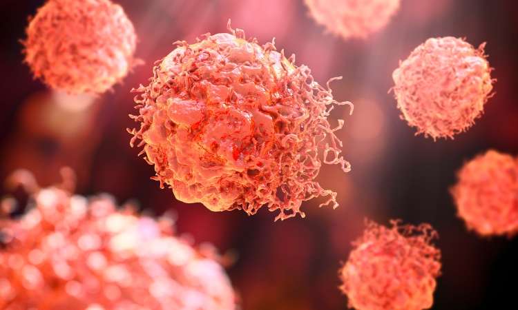

Hosted by Drug Discovery World and supported by Sartorius and BioIVT, this webinar will explore the opportunities and challenges that exist within cancer research drug discovery and development.
You will hear from Dr Sudha Rao, Chief Scientific Officer of Kazia Therapeutics, Karol Budzik, PhD, Business Development Associate at Vyriad Therapeutics and Lars van der Veen, Chief Scientific Officer at iOnctura.
Presentations will cover how cancer treatments have shifted towards reprogramming the biology driving tumour growth, immune escape and treatment resistance, the trajectory that in vivo CAR-T treatments are taking, and how challenging tumours burdened by stroma and immune-mediated resistance can be tackled.
Q&A with the speakers follows the presentations.
The post New webinar: Tackling drug discovery challenges in cancer research appeared first on Drug Discovery World (DDW).
Amid the rise of psychedelics in the mental health space, researchers have begun to explore psilocybin as a treatment for chemotherapy-induced peripheral neuropathy.
Uncategorized
New Spectrometry Technique Could Aid Formulation Development
New Spectrometry Technique Could Aid Formulation Development
A new technique combining two forms of spectrometry could help biopharmaceutical companies improve their choice of formulation buffer for antibody manufacturing by revealing how molecular forms and three-dimensional shapes of complex biologics respond to their environment. That’s the view of Christian Bleiholder, PhD, a professor at Florida State University who helped develop the technique.
According to Bleiholder, what happens structurally when a complex biological molecule, such as an antibody or viral spike protein, binds to its target is currently poorly understood.
“This is where [this approach] can help with the bioprocessing and formulation,” he says, as structural changes “can affect the lifespan [of the product] and lead to issues, such as aggregation.”
Because antibodies are complex, existing techniques tend to be powerful at different levels of complexity, he explains. Mass spectrometry is particularly powerful for distinguishing molecular composition, while structural approaches such as X-ray crystallography and cryo-electron microscopy can provide high-resolution structural information.
The challenge is understanding the link between these things within a heterogeneous sample, he says.
To overcome this, Bleiholder and his team worked with Bruker Daltonics to develop Tandem-Trapped Ion Mobility Spectrometry (Tandem-TIMS). This combines tandem ion mobility spectrometry with tandem mass spectrometry to disentangle three overlapping layers of molecular complexity: molecular form, three-dimensional shape, and binding or assembly state, he says.
He explains that, if the proteins have different structures, they can be characterized with tandem ion mobility spectrometry, and then mass spectrometry can be used to look at their molecular forms and binding states.
Going forward, Bleiholder hopes the technique can be used for formulation development but also earlier, during drug discovery of new products, such as multi-specific antibodies, to determine which molecular states are important and how those change when a biologic engages its target. He also plans to look at automating the technique.
Bleiholder spoke about using Tandem-TIMS at the Bioprocessing Summit in Boston earlier this year.
The post New Spectrometry Technique Could Aid Formulation Development appeared first on GEN – Genetic Engineering and Biotechnology News.


BIO’s plan for ‘securing America’s biotech century’

China’s innovation moment sees a rise in first in class

Windward prices IPO, Rezera postpones its offering: Finance Report

FDA Approves First Drug to Treat Adults with Primary Membranous Nephropathy

How Vancomycin-Resistant Enterococcus Evolved in a Danish Hospital

Cellectis could cut 70% of workforce in shift to in vivo therapies
-

 Uncategorized8 months ago
Novartis buys Synnovation’s PI3Kα inhibitors for $3 billion
-
Endpoints News8 months ago
Novartis to pay $2B upfront to take next-gen PI3Kα inhibitor from Synnovation
-

 Uncategorized6 months ago
Optical Pooled CRISPR Screen Reveals Regulators of NF-κB Dynamics in Human Cells
-
Uncategorized6 months ago
Relay’s PI3Kα inhibitor clears efficacy bar in Phase 2 vascular anomalies study
-
Uncategorized5 months ago
Hybrid solid−liquid optics enable scalable, high-resolution light-sheet microscopy across diverse immersion media
-
Fierce Biotech8 months ago
Scientists turn pig semen extract into eye drops that kill cancer in mice
-
Nature Biotechnology8 months ago
Sustained nitric oxide production by engineered E. coli remodels the tumor microenvironment and potentiates immunotherapy
-

 BioPharma Dive – AI / Data8 months ago
AstraZeneca lung drug gets ‘surprise’ win in COPD trials
 Scan to Read