
Uncategorized
Protein Protects Against Tau Tangles, Synaptic Loss in Mouse Model of Tauopathy
Protein Protects Against Tau Tangles, Synaptic Loss in Mouse Model of Tauopathy

Alzheimer’s disease (AD) and many other forms of neurodegeneration share a common culprit. In these diseases, tau proteins that normally stabilize neuronal microtubule filaments within nervous system networks instead form noxious knots and gradually disrupt the circuits they would otherwise preserve.
Scientists at Sanford Burnham Prebys have now shown that a different protein known as SORLA offers protection against the effects of these lethal loops. The results of the researcher’s’ study in mice suggests that future research may yield new treatments capable of boosting this protein’s ability to defend the brain.
Timothy Huang, PhD, assistant professor in the Center for Neurologic Diseases at Sanford Burnham Prebys, is senior and corresponding author of the team’s published paper in Science Advances, titled “SORLA up-regulation suppresses pathological effects in aged tauopathy mouse brain,” in which they concluded “These findings reveal a protective role for SORLA in multiple aspects of tauopathy pathogenesis and highlight its potential as a therapeutic target.”
Normally, tau proteins are found throughout the brain and nervous system, helping to maintain the shape and structure of our neuronal wiring. But in certain diseases, including Alzheimer’s disease, tau proteins clump together inside nerve cells, forming what are known as tau tangles. These toxic tangles are linked to cognitive impairment and nerve cell death in diseases known as tauopathies. “In AD, amyloid-β (Aβ) plaques and neurofibrillary tangles (NFTs) comprising hyperphosphorylated tau accumulate in brain,” the authors explained.
The new study focused on the safeguarding capabilities of protein known as SORLA. “A role for the trafficking receptor SORLA (Sortilin-related receptor containing LDLR class A repeats) in reducing Aβ levels has been well established,” the investigators continued. “… however, relatively little is known with respect to whether and how SORLA can potentially affect tau pathology in vivo.”
Timothy Huang added, “In the last 15 or 20 years, considerable data has come out from our lab and other groups showing that SORLA can suppress one of the hallmarks of Alzheimer’s disease—amyloid-beta generation and accumulation. Very little was known, however, about whether SORLA affected the tau tangles reflected on the other side of the coin in Alzheimer’s disease.”
SORLA is expressed in both neurons and glia in mouse and human brain, the authors noted. For their newly reported study the team began by crossbreeding mice that produce extra human SORLA protein, with PS19 (P301S) mice that develop tau tangles, brain atrophy and cognitive deficits. This new mouse model enabled experiments to determine SORLA’s effects on tau protein buildup and its resulting harms.
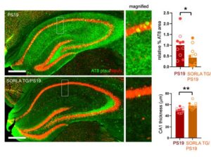
Their studies showed that an overabundance of SORLA protein protected against a number of biological processes linked to the formation of tau tangles and progression of neurodegeneration. These include reducing the addition of too many phosphate groups to tau—known as hyperphosphorylation—and the ability of misshapen tau to serve as “seeds” that attract more tau and form clumps. This protection also extended to preservation of the synapses at the junction between neurons and the brain’s ability to adjust these connection points—which is called synaptic plasticity. “Using complementary approaches, we show that SORLA overexpression attenuates ventricular enlargement, tau phosphorylation and seeding, synaptic loss, impaired synaptic plasticity, and glial hyperactivation in the PS19 mouse brains,” the team wrote in summary.
“When you upregulate SORLA, you can suppress the negative effects found in tauopathies,” said first author Huijie Huang, PhD, a staff scientist in the Huang lab at Sanford Burnham Prebys. “We found there was less brain atrophy and less tau accumulation, which was very exciting to see.”
Because some people have mutations that disable the gene carrying the code for SORLA, Sorl1, the scientists wanted to compare the outcome of having extra SORLA to having none of it at all. Tests of mice genetically modified to lack Sorl1 told a very different story. “The opposite turned out to be true when we deleted the ability to produce SORLA proteins,” said Timothy Huang. “A lack of SORLA exacerbated the harmful effects observed in tauopathies.”
To address how extra SORLA or a lack of SORLA were either ameliorating or aggravating diseases featuring tau tangles, the research team used a combination of sequencing techniques capturing the levels of all proteins and gene expression in each cell, as well as mapping the spatial relationship of RNA and proteins within brain tissue. The scientists found that upregulated SORLA prevented problematic protein production changes in the synapses between neurons while also suppressing other drivers of tauopathy disease progression. They also observed that extra SORLA tamped down on disease-related gene expression patterns in brain cells known as glial cells that support and protect neurons in many ways. “One particularly notable finding that we can build on is the upregulation of a member of the plexin-B family of receptors in the absence of SORLA,” said Huijie Huang.
“There are unique drugs that can target this class of receptors that we may be able to apply to tau-related dementia disorders,” suggested Tim Huang. “One potential future direction is to repurpose these drugs to target overactivation of glial cells and perhaps reverse some of the phenotypes in tauopathies.”
The scientists also want to better understand what happens in each individual cell type when they upregulate or downregulate SORLA. “While it is not possible to specifically determine how cell-specific modulation of SORLA can affect tau using the global transgenic overexpression/deletion models used here, we are interested in further characterizing specific effects of SORLA on tau in neurons, and the extent of SORLA modulation on glia in influencing overall tau pathology,” they stated. The team plans to graft human neurons or glial cells into the mouse brain to study the effects of different SORLA mutations.
“Mouse cells and human cells are different,” said Tim Huang. “Because we’re looking at human disease, it’s more informative if we can observe the modulation and dysfunction of SORLA in the context of a human cell inside of a diseased brain environment.”
This continued research will reveal more knowledge about the ability of SORLA to safeguard against the toxic effects of tau tangles, and how to develop new treatments or repurpose existing therapies to benefit patients suffering from Alzheimer’s disease and other tau-related dementia disorders.
The post Protein Protects Against Tau Tangles, Synaptic Loss in Mouse Model of Tauopathy appeared first on GEN – Genetic Engineering and Biotechnology News.

Post Content
Uncategorized
Capricor CEO won’t rule out legal action against FDA after negative adcomm
Capricor CEO won’t rule out legal action against FDA after negative adcomm
After an FDA advisory committee voted 9-3 on Wednesday against recommending Capricor Therapeutics’ Duchenne muscular dystrophy cardiomyopathy drug for approval, stakeholders allege an “ulterior motive” and “biased intentions.” Read More
During ketamine anesthesia the brain’s nerve cells fall silent, and as consciousness returns, they begin to reconnect. A study in mice by researchers at the Institute of Science and Technology Austria (ISTA) and colleagues at Allen Institute for Brain Science in Seattle, has now for the first time shown that immune cells in the brain play a critical role in this process, and indicates that there are differences between female and male brains.
Senior and corresponding author Sandra Siegert, PhD, at ISTA, reported on their findings in Science Advances, in a paper titled “Corticosterone-linked microglial activity underpins sexually dimorphic neuroplasticity after ketamine anesthesia,” in which they concluded “Our study uncovers significant sex-specific differences in neuronal adaptation during recovery from ketamine anesthesia, driven by microglia.”
Recovery from anesthesia is critical for resuming normal physiological and neuronal functions, but the mechanisms involved remain elusive, the authors wrote. “Ketamine distinguishes itself from other anesthetics by its unique pharmacological properties as an N-methyl D-aspartate (NMDA) receptor antagonist, which preferentially targets GABAergic inhibitory interneurons.”
And unlike many other anesthetics, ketamine does more than induce unconsciousness. It alters how we perceive pain and form memories. It dampens communication between neurons—the very network that must later resume normal function as the patient awakens. Exactly how this recovery process unfolds—and whether male and female brains differ in this regard—has been unclear. “… ketamine anesthesia induces mild anxiety behavior phenotypes, interestingly, only in females,” the team continued, “suggesting inherent sex differences in anesthesia recovery with neuronal consequences that extend beyond the immediate sex-dependent metabolic processing described for low-dose ketamine.”
Working with mice, Siegert, together with Alessandro Venturino, PhD, and their colleagues at ISTA, and researchers at the Allen Institute, now offer the first answers to some of these questions. Microglia are specialized immune cells that constantly scan the brain and, when needed, trigger anti‑inflammatory responses. They also monitor neurons and their connections, thus helping to maintain optimal brain function. “Ketamine, across different dosages, affects microglia, which are embedded within the neuronal network,” the team explained. “Locally, microglia influence the synaptic machinery and neuronal firing properties by responding to environmental changes.”
As early as 2017, Siegert’s group at ISTA noticed that male and female mice respond differently to ketamine anesthesia, and more precisely, their microglia do.
For the newly reported study, using a cranial window—a surgically implanted opening that allows microscopic access to the living brain—Venturino analyzed how microglia and neurons behave while mice recover from ketamine anesthesia. Both cell types were labeled with fluorescent markers to glow under the microscope.
The researchers observed microglia processes in their dynamic action towards neurons. Surprisingly, as female mice recovered from anesthesia, microglia began forming prolonged contacts with neurons, coinciding with the onset of synaptic remodeling and plasticity. Notably, this phenomenon was not observable in male mice.
Furthermore, in mice lacking microglia, no such synaptic remodeling occurred, indicating that microglia are critical mediators of this recovery-associated plasticity. “What was fascinating,” Venturino explains, “was that we observed this plasticity—the brain’s ability to change, adapt, and in this case recover—only in females.”
Despite many other projects—or perhaps because of them—the researchers kept returning to their initial observation. “I’ve always believed that women have greater brain plasticity,” Siegert said with a smile. “Alessandro and I just couldn’t let it go—we wanted to know why.”
Further experiments revealed that this plasticity depends on corticosterone, one of the major stress hormones. “During recovery from anesthesia, corticosterone levels rise,” Venturino explained. “In female mice, this specifically activates the stress‑response gene Fkbp5 in microglia. The gene encodes the protein FKBP51, which helps the cell manage stress signals—and apparently prompts microglia to interact with neurons.”
The authors further noted, “Mechanistically, we found that female microglia selectively up-regulated the cochaperone Fkbp5/FK506-binding protein 51 (FKBP51), which is a key intermediary in the corticosteroid-induced stress response … Our findings underscore that microglia serve as a relevant interface between the endocrine stress response and the brain -immune cell system.”
To confirm this link, the team removed the adrenal glands—the endocrine organs that produce corticosterone. Without them, the close contact between microglia and neurons during recovery disappeared. “These results clearly show that corticosterone triggers this reaction in female mice,” said Venturino.
Siegert added, “They also remind us that stress is not always harmful—stress hormones are essential for certain processes in the brain.” In their paper the investigators commented, “The selective hypothalamic activation and elevated blood plasma corticosterone levels during the recovery phase in females shape the microglia-neuron interactions, highlighting a link between the endocrine and the brain-immune axes.”
Why this process differs between female and male mice remains uncertain; it is still unclear whether the male brain uses a similar mechanism, just delayed, or has another strategy. “Microglia enable rapid adaptation, and these cells in females are likely more sensitive to specific stress signals,” Siegert noted.
From an evolutionary viewpoint, she speculates, females may have faced greater demands for social, emotional, and multitasking adaptability—for example, in childcare, food gathering, or coordinating group activities. The female brain, therefore, had to adapt and respond more swiftly. “That’s a good thing,” Siegert added “But if this plasticity becomes too frequent or too intense, it can increase the risk of depression. We also know that psychiatric disorders are more prevalent in women than in men.”
Siegert further pointed out that during the literature review, her team found very few studies in which ketamine had been tested in females. “There were only a handful of anecdotal studies showing that women experience nausea and sickness more often after ketamine anesthesia,” she stated. Given that ketamine is also used as an antidepressant, understanding how its mechanisms differ between the sexes is all the more important. “It’s astonishing how readily people assume that men and women respond to drugs in the same way—when clearly they do not,” Siegert stressed.
Research like this is a step in the right direction: it highlights that medications can act differently in women and men and serves as a call to consider sex‑specific differences in future studies. “Our findings contribute to a growing body of evidence recognizing sex-specific differences in brain function and immune responses, the latter of which is already well known for increased susceptibility to infection and autoimmune diseases,” the team pointed out. “Our results identified a link between microglia-specific Fkbp5 expression and ketamine action, warranting a reevaluation of assumptions that ketamine is a general anesthetic and fully reversible across sexes.”
Insights into the microglia response to ketamine have since inspired Siegert and Venturino to co‑found Syntropic Medical, a start‑up in ISTA’s XISTA ecosystem exploring how 60 Hz flickering light can soften such neural networks in the brain.
The post Ketamine Triggers Sex-Specific Brain Recovery Responses appeared first on GEN – Genetic Engineering and Biotechnology News.


Illinois’ financial crisis could bring the state to a halt
The final 6 ‘Game of Thrones’ episodes might feel like a full season
New Season 8 Walking Dead trailer flashes forward in time
Mod turns ‘Counter-Strike’ into a ‘Tekken’ clone with fighting chickens
Meet Superman’s grandfather in new trailer for Krypton
Disney’s live-action Aladdin finally finds its stars
De novo L-(+)-tartaric acid biosynthesis in multi-modular engineered yeasts
Capricor CEO won’t rule out legal action against FDA after negative adcomm
Ketamine Triggers Sex-Specific Brain Recovery Responses

STAT+: Trump administration revises rebate pilot for 340B drug discount program, angering hospitals
Microbiota-Derived Metabolite Enhances HIV Therapy in Monkeys
Lilly and Resilience invest $750M in diabetes and obesity drug device supply
Illinois’ financial crisis could bring the state to a halt
The final 6 ‘Game of Thrones’ episodes might feel like a full season
New Season 8 Walking Dead trailer flashes forward in time
Mod turns ‘Counter-Strike’ into a ‘Tekken’ clone with fighting chickens
Meet Superman’s grandfather in new trailer for Krypton
Disney’s live-action Aladdin finally finds its stars
-
Fierce Biotech4 months ago
Scientists turn pig semen extract into eye drops that kill cancer in mice
-
 Uncategorized9 years ago
These ’90s fashion trends are making a comeback in 2017
-
Endpoints News4 months ago
Novartis to pay $2B upfront to take next-gen PI3Kα inhibitor from Synnovation
-

 Uncategorized4 months ago
Novartis buys Synnovation’s PI3Kα inhibitors for $3 billion
-
Uncategorized9 years ago
According to Dior Couture, this taboo fashion accessory is back
-
Uncategorized9 years ago
Phillies’ Aaron Altherr makes mind-boggling barehanded play
-
Uncategorized9 years ago
Steph Curry finally got the contract he deserves from the Warriors
-
Uncategorized9 years ago
Uber and Lyft are finally available in all of New York State