

Uncategorized
Personalized Cartilage Graft Developed for Life-Threatening Infant Airway Narrowing

A study led by researchers at Children’s Hospital of Philadelphia (CHOP) demonstrated a new method of using decellularized cartilage with patient-specific cells to help enlarge the pediatric airways narrowed as a result of severe subglottic stenosis (SGS). The condition is a narrowing of the airway below the vocal cords and above the trachea, and affects an estimated 20,000 infants per year.
Researchers demonstrated in a preclinical model that this new method of airway reconstruction was faster, more effective, and able to overcome issues, such as donor site morbidity, insufficient tissue volume and delayed timeline, associated with the current standard grafts used for laryngotracheal reconstruction (LTR).
Riccardo Gottardi, PhD, assistant professor with the Perelman School of Medicine at the University of Pennsylvania and leader of the Bioengineering and Biomaterials (Bio2) lab, and Ian Jacobs, MD, medical director of the Center for Pediatric Airway Disorders in the Division of Otolaryngology (ENT) at CHOP, co-led the research, which is reported in Nature Communications, in a paper titled “A translational approach to airway reconstruction leveraging decellularized meniscus and cartilage progenitor cells.” In their paper the team said, “This technology has the potential to revolutionize the field of pediatric LTR.”
Severe subglottic stenosis (SGS) develops in children almost exclusively as a response to intubation, and affects nearly 1.5% of the over 200,000 infants in intensive care units each year in the United States, the authors explained. The most severe cases require laryngotracheal reconstruction (LTR), an open airway surgery that is used to enlarge the airway by implanting cartilage taken from a rib cage. While LTR is used to successfully treat thousands of children with subglottic stenosis, in many cases, young children often lack enough costal cartilage—the cartilage connecting our ribs to the sternum—for these grafts.
As a result, operations often need to be delayed, leaving the child attached to a tracheostomy tube until they are older and grown enough to supply sufficiently sized cartilage for an effective LTR, and there is a higher risk of needing follow-up surgery because the airway is at risk of narrowing again. “In adults, LTR has a 90% success rate with low rates of revision,” the team stated. “However, in children, success rates significantly drop, and the incidence of restenosis requiring revision surgery increases to over 24%.”
To improve this process and reduce the risk of these potential complications, Gottardi and Jacobs and colleagues have been looking at tissue engineering a laryngotracheal graft. Tissue engineering, the team wrote, could provide “an ideal alternative to autologous cartilage grafts to alleviate unnecessary comorbidities as well as reduce surgical time.” However, the complexity of the trachea prevents the use of conventional cartilage engineering techniques for this procedure.
“We needed something that could be equivalent to a piece of cartilage, integrate well with the surrounding tissue, be well tolerated by the patient, behave like native tissues and regrow and be part of the airway,” Gottardi said. “This required quite a bit of creative thinking because of the additional challenges in children who are so small and still growing.”
To overcome the limitations of existing methods, the researchers, led by former Gottardi lab member Paul Gehret, PhD, created a first-of-its-kind scaffold based on porcine meniscal cartilage decellularization (MEND – MENiscus Decellularization). They realized that if the cells, elastin, and blood vessels present in the meniscus are “digested” away, the meniscal cartilage becomes amenable to recellularization and integration while being less likely to provoke an immune response. In their paper the researchers explained, “Building on the strengths of previous decellularized therapies, we established an innovative approach that leverages the selective enzymatic removal of the elastin fibers and blood vessels uniquely present in the fibro-elastic cartilage of the meniscus to create microchannels, which support cellular invasion while substantially preserving native structure.”
Using ear-derived cartilage progenitor cells (eCPCs), which can mature into cartilage-producing chondrocytes, the researchers demonstrated that MEND can be recellularized after the removal of elastin and blood vessels and suitable for implantation in less than a month. “Notably, porcine menisci, such as those used in this study, are a highly abundant cartilage source, being easily available as a waste product of the food industry, which can ensure no shortage of material for surgeons to shape into an ideal implant,” the team noted.
Importantly, the new method needed to work in a clinically relevant timeframe. In a real-world scenario, clinicians may only have one or two months to be able to perform the procedure when it can still benefit the patient. Harvesting seed cells within days and creating a scaffold within three to four weeks is significantly less time than the six months that was typically needed for engineered cartilage. “Here we demonstrate that MEND can be fully recellularized in three days with ear-derived cartilage progenitor cells and reaches structural and functional maturation suitable for implant within three weeks of chondrogenic differentiation, a time frame compatible with clinical translation,” the authors stated.
They validated their technology in a preclinical rabbit in vivo model, and demonstrated better performance than costal cartilage, the standard of care, with no instances of adverse events reported. “Our results demonstrate airway expansion, graft reepithelialization, neocartilage formation, and integration with adjacent native laryngotracheal cartilage at three months,” the team stated. “Notably, MEND implants perform better in all outcomes than autologous costal cartilage, the standard of care.”
These findings will be further validated prior to proposing the procedure for patients suffering from severe subglottic stenosis. “These results demonstrate the feasibility of our translational tissue engineering approach to laryngotracheal reconstruction and could overcome the autograft-associated limitations in pediatric patients, decreasing the need for invasive revision surgery,” the investigators concluded.
“This research shows really promising data that suggests this novel approach could overcome the autograft-associated limitations we sometimes encounter when attempting laryngotracheal reconstruction in infants,” Jacobs said. “With more research, we expect this could decrease the need for invasive surgery, and we may be able to apply the technology to other conditions that require a cartilage graft.”
The post Personalized Cartilage Graft Developed for Life-Threatening Infant Airway Narrowing appeared first on GEN – Genetic Engineering and Biotechnology News.

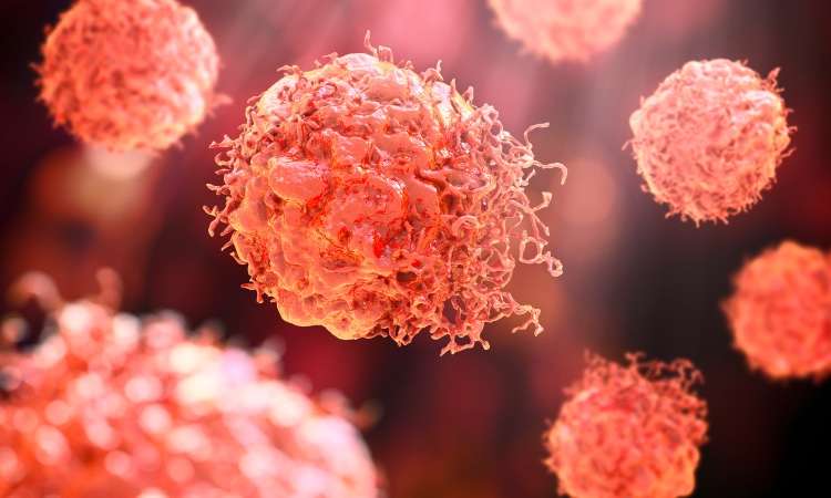

Hosted by Drug Discovery World and supported by Sartorius and BioIVT, this webinar will explore the opportunities and challenges that exist within cancer research drug discovery and development.
You will hear from Dr Sudha Rao, Chief Scientific Officer of Kazia Therapeutics, Karol Budzik, PhD, Business Development Associate at Vyriad Therapeutics and Lars van der Veen, Chief Scientific Officer at iOnctura.
Presentations will cover how cancer treatments have shifted towards reprogramming the biology driving tumour growth, immune escape and treatment resistance, the trajectory that in vivo CAR-T treatments are taking, and how challenging tumours burdened by stroma and immune-mediated resistance can be tackled.
Q&A with the speakers follows the presentations.
The post New webinar: Tackling drug discovery challenges in cancer research appeared first on Drug Discovery World (DDW).
Amid the rise of psychedelics in the mental health space, researchers have begun to explore psilocybin as a treatment for chemotherapy-induced peripheral neuropathy.
Uncategorized
New Spectrometry Technique Could Aid Formulation Development
New Spectrometry Technique Could Aid Formulation Development
A new technique combining two forms of spectrometry could help biopharmaceutical companies improve their choice of formulation buffer for antibody manufacturing by revealing how molecular forms and three-dimensional shapes of complex biologics respond to their environment. That’s the view of Christian Bleiholder, PhD, a professor at Florida State University who helped develop the technique.
According to Bleiholder, what happens structurally when a complex biological molecule, such as an antibody or viral spike protein, binds to its target is currently poorly understood.
“This is where [this approach] can help with the bioprocessing and formulation,” he says, as structural changes “can affect the lifespan [of the product] and lead to issues, such as aggregation.”
Because antibodies are complex, existing techniques tend to be powerful at different levels of complexity, he explains. Mass spectrometry is particularly powerful for distinguishing molecular composition, while structural approaches such as X-ray crystallography and cryo-electron microscopy can provide high-resolution structural information.
The challenge is understanding the link between these things within a heterogeneous sample, he says.
To overcome this, Bleiholder and his team worked with Bruker Daltonics to develop Tandem-Trapped Ion Mobility Spectrometry (Tandem-TIMS). This combines tandem ion mobility spectrometry with tandem mass spectrometry to disentangle three overlapping layers of molecular complexity: molecular form, three-dimensional shape, and binding or assembly state, he says.
He explains that, if the proteins have different structures, they can be characterized with tandem ion mobility spectrometry, and then mass spectrometry can be used to look at their molecular forms and binding states.
Going forward, Bleiholder hopes the technique can be used for formulation development but also earlier, during drug discovery of new products, such as multi-specific antibodies, to determine which molecular states are important and how those change when a biologic engages its target. He also plans to look at automating the technique.
Bleiholder spoke about using Tandem-TIMS at the Bioprocessing Summit in Boston earlier this year.
The post New Spectrometry Technique Could Aid Formulation Development appeared first on GEN – Genetic Engineering and Biotechnology News.


Activities Report of the Generic Drug Program | FDARA Title VIII Sections 807 and 805

BIO’s plan for ‘securing America’s biotech century’

China’s innovation moment sees a rise in first in class

Windward prices IPO, Rezera postpones its offering: Finance Report

FDA Approves First Drug to Treat Adults with Primary Membranous Nephropathy

How Vancomycin-Resistant Enterococcus Evolved in a Danish Hospital
-

 Uncategorized8 months ago
Novartis buys Synnovation’s PI3Kα inhibitors for $3 billion
-
Endpoints News8 months ago
Novartis to pay $2B upfront to take next-gen PI3Kα inhibitor from Synnovation
-
 Uncategorized6 months ago
Optical Pooled CRISPR Screen Reveals Regulators of NF-κB Dynamics in Human Cells
-
Uncategorized6 months ago
Relay’s PI3Kα inhibitor clears efficacy bar in Phase 2 vascular anomalies study
-
Uncategorized5 months ago
Hybrid solid−liquid optics enable scalable, high-resolution light-sheet microscopy across diverse immersion media
-
Fierce Biotech8 months ago
Scientists turn pig semen extract into eye drops that kill cancer in mice
-
Nature Biotechnology8 months ago
Sustained nitric oxide production by engineered E. coli remodels the tumor microenvironment and potentiates immunotherapy
-

 BioPharma Dive – AI / Data8 months ago
AstraZeneca lung drug gets ‘surprise’ win in COPD trials
 Scan to Read