

Uncategorized
iPSC-Derived Retinal Endothelial Cells Offer Platform for Studying Diseases
iPSC-Derived Retinal Endothelial Cells Offer Platform for Studying Diseases

Biomedical engineers at Duke University have for the first time used induced pluripotent stem cells (iPSCs) to grow specialized blood vessel cells critical to retinal health.
When injected into mouse models of retinal disease, these “retinal endothelial cells” (iRECs) integrated into the damaged tissue to regenerate blood vessels and restore retinal function. The team also demonstrated these cells’ ability to form functional retinal vascular tissue in a lab-grown environment, providing a pathway to model and research various eye diseases.
The results point toward the potential of using these retinal cells and models to develop new methods of impactful vision loss treatments and eye disorder research. “Retinal vascular diseases affect millions of people in the U.S., but our understanding remains limited, hindering our ability to discover and develop new therapeutics,” said Sharon Gerecht, PhD, the Paul M. Gross Distinguished professor and chair of Biomedical Engineering at Duke. “Using human stem cells, we generated the cells found in retinal blood vessels, paving the way for new therapeutic approaches.”
Gerecht is senior and corresponding author of the researchers’ published paper in Nature Biomedical Engineering, titled “Derivation of functional retinal endothelial cells from human pluripotent stem cells for therapeutics and modeling.” In their report the authors suggested that their iREC differentiation strategy will “… advance cell therapy and disease modeling, accelerating the discovery of treatments for retinal microvascular diseases.”
The old saying that the eyes are windows into the soul is more accurate than one might think. Neurons from the retina—the back part of the eye that detects light—extend directly to the brain, technically making the eyes part of the central nervous system.
Also like the brain, the retina has a blood barrier that strictly controls what gets in and out including oxygen, nutrients, water and pharmaceuticals. While this barrier keeps the retina healthy and relatively protected from disease-causing agents, it also makes treating the retina difficult. “Retinal tissue has the highest energy and oxygen usage in the body due to the retina’s intense and continuous neuronal activity,” the authors further explained. “This demand leads to a crucial reliance on the inner blood–retina barrier (iBRB) to maintain ocular homeostasis.”
The barrier is formed by blood vessel tissue comprising a tight network of retinal endothelial cells, which form the inner layer of blood vessels, in concert with other specialized cells called pericytes and astrocytes. “Retinal endothelial cells (RECs) in the iBRB are continuous endothelial cells (ECs) that form tight junctions to regulate the diffusion of small molecules, such as ions and water, across their cell–cell interface,” the investigators continued. The specificity of these cells and the fact that they do not form in other areas of the body make the complex tissue difficult to heal or to grow from scratch.
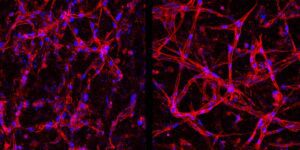
“When this specialized blood vessel tissue begins to break down, it can cause a lot of different diseases that lead to vision loss,” said Parker Esswein, a PhD student working in the Gerecht laboratory and co-first author of the paper. “While there are sources of retinal endothelial cells, being able to grow a continuous supply from scratch could offer many advantages for those working in the field.”
These retinal endothelial cells are currently collected and grown from real patients, making them relatively expensive with a limited supply. “A renewable source of human iBRB endothelium is thus vital for advancing eye research and treatment development,” the team noted in their paper.
To expand access, reduce cost and control variability, the Gerecht lab wanted to see if they could grow them from iPSCs. These are essentially mature adult cells reprogrammed to become primal versions of themselves that can then grow into a wide variety of other cell types.
To do this, Esswein and Ying-Yu Lin, PhD, a former PhD student in Gerecht’s lab, took commercial iPSCs and used a well-established procedure to get them to grow into common endothelial cells that form the inner layer of most of the body’s blood vessels. The researchers then used a specialized cocktail of growth factors to coax the cells into becoming the specific type of endothelial cells found in the retina. “… we differentiated human induced pluripotent stem cells into retinal endothelial cells (iRECs) via the Wnt–β-catenin pathway, namely Norrin–Frizzled4 signaling,” they explained.
Once successful, the researchers put their development to the test. In benchtop experiments, the team was able to get the iRECs to form the same networks and structures that they do within the body. The team then subjected these lab-grown tissues to low oxygen and high glucose levels, which are detrimental conditions often seen within real people. These conditions are fundamental causes of diabetic retinopathy (DR), the leading cause of vision loss in working-age people in the United States, and caused the tissue barrier to break down just like it does in patients. They wrote in summary, “Overall, we were able to robustly recapitulate the DR phenotype in 2D and 3D with our iRECs, exemplifying their ability to be utilized for in vitro disease modeling and to elucidate aberrant pathways and therapeutic targets.”
The researchers then tried their lab-grown cells as a therapy for mouse models with weak, unstructured retinal blood vessels. When injected into the mice before any actual vision loss occurred, these cells successfully integrated into the existing tissue and helped develop strong blood vessels with strong barriers. “When injected into oxygen-induced retinopathy mice, iRECs integrated into the host vascular network and revascularized the ischemic eye, rescuing the tissue,” they stated.
“The tests showed that these lab-grown cells have promise for preventative treatments, especially since they should be easier and cheaper to obtain using our technique,” Esswein said. “And while our benchtop experiments did not attempt to model a wide variety of specific eye diseases in these studies, we’re confident we can create excellent human tissue models in the lab to help better understand these diseases and uncover therapies.”
Moving forward, the researchers are planning to explore these potential uses for their retinal endothelial cells both in their laboratory and through emerging industry partnerships. The group also has a patent pending that covers both the stem cell-based therapeutics and in vitro modeling for drug discovery and testing. In their paper they concluded “Our study establishes functional human iRECs and microphysiological iBRB models that facilitate mechanistic studies aimed at identifying therapeutic targets and promoting the revascularization of injured retinas, thereby supporting treatment advancement.”
The post iPSC-Derived Retinal Endothelial Cells Offer Platform for Studying Diseases appeared first on GEN – Genetic Engineering and Biotechnology News.


Hosted by Drug Discovery World and supported by Sartorius and BioIVT, this webinar will explore the opportunities and challenges that exist within cancer research drug discovery and development.
You will hear from Dr Sudha Rao, Chief Scientific Officer of Kazia Therapeutics, Karol Budzik, PhD, Business Development Associate at Vyriad Therapeutics and Lars van der Veen, Chief Scientific Officer at iOnctura.
Presentations will cover how cancer treatments have shifted towards reprogramming the biology driving tumour growth, immune escape and treatment resistance, the trajectory that in vivo CAR-T treatments are taking, and how challenging tumours burdened by stroma and immune-mediated resistance can be tackled.
Q&A with the speakers follows the presentations.
The post New webinar: Tackling drug discovery challenges in cancer research appeared first on Drug Discovery World (DDW).
Amid the rise of psychedelics in the mental health space, researchers have begun to explore psilocybin as a treatment for chemotherapy-induced peripheral neuropathy.
Uncategorized
New Spectrometry Technique Could Aid Formulation Development
New Spectrometry Technique Could Aid Formulation Development
A new technique combining two forms of spectrometry could help biopharmaceutical companies improve their choice of formulation buffer for antibody manufacturing by revealing how molecular forms and three-dimensional shapes of complex biologics respond to their environment. That’s the view of Christian Bleiholder, PhD, a professor at Florida State University who helped develop the technique.
According to Bleiholder, what happens structurally when a complex biological molecule, such as an antibody or viral spike protein, binds to its target is currently poorly understood.
“This is where [this approach] can help with the bioprocessing and formulation,” he says, as structural changes “can affect the lifespan [of the product] and lead to issues, such as aggregation.”
Because antibodies are complex, existing techniques tend to be powerful at different levels of complexity, he explains. Mass spectrometry is particularly powerful for distinguishing molecular composition, while structural approaches such as X-ray crystallography and cryo-electron microscopy can provide high-resolution structural information.
The challenge is understanding the link between these things within a heterogeneous sample, he says.
To overcome this, Bleiholder and his team worked with Bruker Daltonics to develop Tandem-Trapped Ion Mobility Spectrometry (Tandem-TIMS). This combines tandem ion mobility spectrometry with tandem mass spectrometry to disentangle three overlapping layers of molecular complexity: molecular form, three-dimensional shape, and binding or assembly state, he says.
He explains that, if the proteins have different structures, they can be characterized with tandem ion mobility spectrometry, and then mass spectrometry can be used to look at their molecular forms and binding states.
Going forward, Bleiholder hopes the technique can be used for formulation development but also earlier, during drug discovery of new products, such as multi-specific antibodies, to determine which molecular states are important and how those change when a biologic engages its target. He also plans to look at automating the technique.
Bleiholder spoke about using Tandem-TIMS at the Bioprocessing Summit in Boston earlier this year.
The post New Spectrometry Technique Could Aid Formulation Development appeared first on GEN – Genetic Engineering and Biotechnology News.


STAT+: FDA approves new breast cancer drug, providing new option for some patients, boost for Roche

Continuous Pediatric Antibiotic Infusions Reduce Preparation Time Without Altering Perceived Workload

Mendelian Randomization Study Links Specific Viral Antibodies to Prostate Cancer Risk

Review Evaluates Integrin Alpha-9-Beta-1 Potential in Cancer Theranostics and Preclinical Limits

New Chemotherapy Quality of Life Scale Validated for Breast Cancer Patients

Low Decisional Conflict Among Prostate Cancer Patients Seeking Germline Testing
-

 Uncategorized8 months ago
Novartis buys Synnovation’s PI3Kα inhibitors for $3 billion
-
Endpoints News8 months ago
Novartis to pay $2B upfront to take next-gen PI3Kα inhibitor from Synnovation
-

 Uncategorized7 months ago
Optical Pooled CRISPR Screen Reveals Regulators of NF-κB Dynamics in Human Cells
-
Uncategorized6 months ago
Relay’s PI3Kα inhibitor clears efficacy bar in Phase 2 vascular anomalies study
-
Uncategorized5 months ago
Hybrid solid−liquid optics enable scalable, high-resolution light-sheet microscopy across diverse immersion media
-
Fierce Biotech8 months ago
Scientists turn pig semen extract into eye drops that kill cancer in mice
-
Nature Biotechnology8 months ago
Sustained nitric oxide production by engineered E. coli remodels the tumor microenvironment and potentiates immunotherapy
-

 BioPharma Dive – AI / Data8 months ago
AstraZeneca lung drug gets ‘surprise’ win in COPD trials
 Scan to Read