

Uncategorized
In Huntington’s Mouse, Optogenetic Activation of VIP Neurons Restores Brain Function
In Huntington’s Mouse, Optogenetic Activation of VIP Neurons Restores Brain Function

Huntington’s disease is a devastating brain disorder in which damage to nerve cells leads to progressively worsening cognitive and movement abilities. While the genetic mutation responsible for the condition is well known, the details of how the disease disrupts brain circuits have not been clearly understood. Now, researchers have identified and tracked neurons involved in Huntington’s disease progression and used optogenetics to selectively activate these neurons and improve the debilitating deficits of the condition.
The study is published in Nature in the paper, “Restoring cortical disinhibition improves Huntington’s disease phenotypes.”
“This work shows that correcting specific imbalances in brain circuits can restore function, even in a complex neurodegenerative condition, and highlights the potential of targeting defined cell types to promote recovery,” said Takaki Komiyama, PhD, professor in the UC San Diego Departments of Neurobiology (School of Biological Sciences) and Neurosciences (School of Medicine).
Huntington’s disease is caused by a trinucleotide repeat mutation in the Huntingtin (HTT) gene. While the mutation is well known, the neural networks connected with the disease progression have been more elusive.
This work aimed to map the neural circuits that expose the networks involved at the onset and spread of the disease’s debilitating symptoms. In transgenic mice carrying the same mutation as human patients, the researchers evaluated how different types of brain cells in the motor cortex are affected in Huntington’s disease. Advanced imaging techniques allowed the researchers to track the activity of these cortical neurons as the disorder progressed.
The researchers found that the disease disrupts the balance of activity across different cell types, including cortical inhibitory neurons.
“Cortical inhibitory cells have received little attention in Huntington’s disease, as for a long time they were considered to be spared from neurodegeneration,” said Irina Dudanova, PhD, previously based at the Max Planck Institute for Biological Intelligence, now at the University of Würzburg in Germany. “Surprisingly, we detected profound changes in their activity, with some cell types being overactive and some nearly silent.”
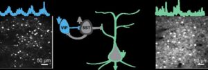
In particular, a class of inhibitory neurons known as vasoactive intestinal peptide (VIP) neurons, exhibited significantly reduced activity. VIP neuron activity is essential for normal learning, as these cells enable the brain to adapt and refine brain circuits during learning.
Reduced VIP neuron activity, the researchers reasoned, could be impairing the brain’s ability to function and learn properly. They sought to activate these cells to re-engage brain states that support learning. They tested this idea using optogenetics to stimulate VIP neurons.
“By activating the VIP inhibitory cell type, we gradually restored more normal activity patterns, and, very importantly, we also saw an improvement in the ability of the mouse to learn a motor task,” said Sonja Blumenstock, PhD, assistant project scientist at UC San Diego.
The results confirm VIP neurons as a key point of vulnerability in Huntington’s disease as well as a promising target for therapy. As to how this process works, the results suggest that modulating VIP neurons opens a “gate” that enables learning-related brain plasticity.
“This intervention restored more normal patterns of activity in the brain and improved movement in affected mice,” said Komiyama. “Importantly, the improvements persisted for days after stimulation ended, suggesting that the treatment triggered lasting beneficial changes in brain circuits rather than only temporary effects.”
The study provides important indications of where research could focus to normalize human brain function and facilitate brain recovery. Komiyama envisions a future scenario in which scientists could non-invasively activate the brain from outside the skull using novel approaches.
“Our study shows that despite the genetic defect, a precise intervention into the brain circuitry can lead to significant improvements in motor symptoms,” said Dudanova. “If we know which cells to target, we can retune the brain’s abnormal activity patterns. This gives hope for future therapies.”
The research also shows that corrections to specific brain circuit imbalances can restore function in a highly complex neurodegenerative condition, with similar potential in other disorders.
“We have come up with a way to allow the diseased brain to learn better,” said Komiyama. “The approach can improve behavior in diseased mice, and our hope is that a related approach will help people with impairment in their learning abilities.”
The post In Huntington’s Mouse, Optogenetic Activation of VIP Neurons Restores Brain Function appeared first on GEN – Genetic Engineering and Biotechnology News.


Hosted by Drug Discovery World and supported by Sartorius and BioIVT, this webinar will explore the opportunities and challenges that exist within cancer research drug discovery and development.
You will hear from Dr Sudha Rao, Chief Scientific Officer of Kazia Therapeutics, Karol Budzik, PhD, Business Development Associate at Vyriad Therapeutics and Lars van der Veen, Chief Scientific Officer at iOnctura.
Presentations will cover how cancer treatments have shifted towards reprogramming the biology driving tumour growth, immune escape and treatment resistance, the trajectory that in vivo CAR-T treatments are taking, and how challenging tumours burdened by stroma and immune-mediated resistance can be tackled.
Q&A with the speakers follows the presentations.
The post New webinar: Tackling drug discovery challenges in cancer research appeared first on Drug Discovery World (DDW).
Amid the rise of psychedelics in the mental health space, researchers have begun to explore psilocybin as a treatment for chemotherapy-induced peripheral neuropathy.
Uncategorized
New Spectrometry Technique Could Aid Formulation Development
New Spectrometry Technique Could Aid Formulation Development
A new technique combining two forms of spectrometry could help biopharmaceutical companies improve their choice of formulation buffer for antibody manufacturing by revealing how molecular forms and three-dimensional shapes of complex biologics respond to their environment. That’s the view of Christian Bleiholder, PhD, a professor at Florida State University who helped develop the technique.
According to Bleiholder, what happens structurally when a complex biological molecule, such as an antibody or viral spike protein, binds to its target is currently poorly understood.
“This is where [this approach] can help with the bioprocessing and formulation,” he says, as structural changes “can affect the lifespan [of the product] and lead to issues, such as aggregation.”
Because antibodies are complex, existing techniques tend to be powerful at different levels of complexity, he explains. Mass spectrometry is particularly powerful for distinguishing molecular composition, while structural approaches such as X-ray crystallography and cryo-electron microscopy can provide high-resolution structural information.
The challenge is understanding the link between these things within a heterogeneous sample, he says.
To overcome this, Bleiholder and his team worked with Bruker Daltonics to develop Tandem-Trapped Ion Mobility Spectrometry (Tandem-TIMS). This combines tandem ion mobility spectrometry with tandem mass spectrometry to disentangle three overlapping layers of molecular complexity: molecular form, three-dimensional shape, and binding or assembly state, he says.
He explains that, if the proteins have different structures, they can be characterized with tandem ion mobility spectrometry, and then mass spectrometry can be used to look at their molecular forms and binding states.
Going forward, Bleiholder hopes the technique can be used for formulation development but also earlier, during drug discovery of new products, such as multi-specific antibodies, to determine which molecular states are important and how those change when a biologic engages its target. He also plans to look at automating the technique.
Bleiholder spoke about using Tandem-TIMS at the Bioprocessing Summit in Boston earlier this year.
The post New Spectrometry Technique Could Aid Formulation Development appeared first on GEN – Genetic Engineering and Biotechnology News.


STAT+: FDA approves new breast cancer drug, providing new option for some patients, boost for Roche

Continuous Pediatric Antibiotic Infusions Reduce Preparation Time Without Altering Perceived Workload

Mendelian Randomization Study Links Specific Viral Antibodies to Prostate Cancer Risk

Review Evaluates Integrin Alpha-9-Beta-1 Potential in Cancer Theranostics and Preclinical Limits

New Chemotherapy Quality of Life Scale Validated for Breast Cancer Patients

Low Decisional Conflict Among Prostate Cancer Patients Seeking Germline Testing
-

 Uncategorized8 months ago
Novartis buys Synnovation’s PI3Kα inhibitors for $3 billion
-
Endpoints News8 months ago
Novartis to pay $2B upfront to take next-gen PI3Kα inhibitor from Synnovation
-

 Uncategorized7 months ago
Optical Pooled CRISPR Screen Reveals Regulators of NF-κB Dynamics in Human Cells
-
Uncategorized6 months ago
Relay’s PI3Kα inhibitor clears efficacy bar in Phase 2 vascular anomalies study
-
Uncategorized5 months ago
Hybrid solid−liquid optics enable scalable, high-resolution light-sheet microscopy across diverse immersion media
-
Fierce Biotech8 months ago
Scientists turn pig semen extract into eye drops that kill cancer in mice
-
Nature Biotechnology8 months ago
Sustained nitric oxide production by engineered E. coli remodels the tumor microenvironment and potentiates immunotherapy
-

 BioPharma Dive – AI / Data8 months ago
AstraZeneca lung drug gets ‘surprise’ win in COPD trials
 Scan to Read