
Uncategorized
From monolayer to microtissue: Measuring what matters in 3D drug discovery

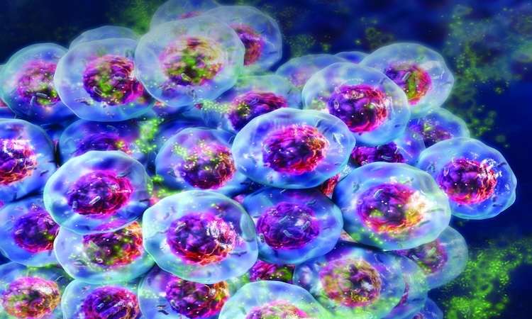

Darren Heywood, Senior Product Manager at Promega UK, examines assay formats built specifically for 3D measurements.
A 3D microtissue, such as a spheroid or organoid, is not a monolayer with more cells. These are three-dimensional cultures that self-organise into structures resembling real tissue. In a spheroid, as it grows, oxygen and nutrients become limited towards the centre, creating a gradient from actively proliferating cells at the outer edge to quiescent, hard-to-reach cells at the core. Those core cells are, in many respects, the ones that matter most. An assay that reads only the outer rim isn’t measuring the wrong model, it’s measuring the wrong cells.
That biology is different from monolayer cultures, and in drug discovery it matters. Potency data from the outer layers reflects the cells most directly exposed to drug, not the resistant, quiescent population at the core that drives relapse and clinical failure. It’s a key reason why compounds that look promising in the lab often disappoint in the clinic.
Endpoint assays: Adapted for the biology
Endpoint assays are the backbone of early compound screening, generating IC50 values and dose-response curves that determine which compounds progress. Running them in 3D is not always straightforward. The most common issue is lysis: standard buffers were designed for flat monolayers, not for cells embedded in matrix, and incomplete lysis means IC50 values are underestimated, reflecting only the most accessible cells rather than the full population. Stronger lysis conditions fix this. CellTiter-Glo 3D was reformulated for this purpose, giving reliable IC50 values from the full depth of the spheroid. The IC50 values are typically higher than in monolayer cultures, not because the assay is less sensitive, but because the spheroid contains quiescent, drug-resistant cells in the hypoxic core that monolayer cultures do not. Caspase-Glo 3/7 3D applies the same enhanced lysis principle to mechanism of action studies, measuring caspase-3/7 activity across the full spheroid depth. In 3D, this readout is more stringent than in 2D. Cells in the hypoxic core actively resist apoptosis through upregulation of anti-apoptotic proteins such as BCL-2 and survivin, a defence mechanism driven by those same oxygen and nutrient gradients. A compound that activates caspase-3/7 in a spheroid is overcoming the hypoxia-driven apoptosis resistance that monolayer cultures do not replicate.
Understanding whether a compound is killing cells or just stopping them from dividing matters for dosing, combination therapy and the likelihood of resistance. The standard marker for this is Ki-67, a nuclear protein expressed only in cells that are actively cycling and absent in quiescent ones. Conventional Ki-67 measurement requires cell fixation, dissociation or sectioning, followed by microscopy or flow cytometry, none of which works at the throughput a compound screen demands. The Lumit Ki-67 assay sidesteps this by detecting Ki-67 from cell lysates in a standard plate reader format with no fixation, no washing and no specialist equipment, making Ki-67 practical for compound screening.
Real-time kinetic assays: Beyond what endpoints can show
Endpoint assays give a snapshot of compound activity at a single point in time. What they cannot show is when those effects start, how fast they develop or whether they reverse. This shapes dosing, scheduling and combination decisions. Real-time kinetic assays add reagents to the culture medium without lysing the cells. The 3D structure stays intact and the same well can be read repeatedly, capturing the full-time course of compound activity.
Kinetic data from 2D cultures does not always translate to 3D. Without the diffusion barriers, quiescent subpopulations and hypoxic gradients that develop in a spheroid, monolayer cells do not present the same resistance mechanisms. Compounds that appear highly active in 2D can show less effect in spheroids of the same cell line. Using the RealTime-Glo Annexin V Apoptosis and Necrosis Assay, Kota et al. showed that Proscillaridin A induced apoptosis at earlier timepoints and at higher rates in oncogenic KRAS spheroids compared with wild-type. This selectivity was completely absent in 2D, where both cell lines were equally inhibited and the compound would have been discarded (Oncogene, 2018). It shows how 3D kinetic measurement can change which compounds advance. What real-time kinetic assays cannot access is what cells release into the medium, and for that a different approach is needed.
Media-sampling assays: A window into biology that other formats miss
Media-sampling assays address this by measuring analytes from the culture medium without disturbing the cells, from the same well, throughout the treatment period.
For metabolic profiling studies, 2D data can overestimate compound activity because the biology that drives metabolic resistance in tumours does not exist in monolayer cultures. Oxygen and nutrient gradients in spheroids drive a Warburg-like glycolytic shift, with elevated glucose consumption and increased lactate secretion, that closely mirrors the tumour microenvironment. In monolayer cultures, cells are uniformly oxygenated and proliferating, so the metabolic readout has little relevance to what a compound encounters in vivo. Glucose-Glo and Lactate-Glo measure these outputs from as little as 2–5µL of conditioned media, leaving the spheroid intact. Glutamine adds a further dimension: quiescent cells in the hypoxic core are less glutamine-dependent than actively cycling cells, so glutaminase inhibitors appear more potent in 2D than in 3D. Glutamine/Glutamate-Glo picks this up early, before it becomes a clinical problem.
CYP450 activity illustrates what 3D adds for safety assessment. Primary hepatocytes in monolayer lose CYP activity within 24 to 48 hours, often before the experiment is finished. The same cells in 3D microtissues retain it for days to weeks, making drug-drug interaction and metabolic liability studies reliable in a way that 2D cannot. P450-Glo Assays measure CYP activity directly from the culture medium, so the same microtissue can be followed across the full study.
Multiplexing: The complete picture from one well
In 2D, running separate assays on separate wells is straightforward as cells are plentiful and easily replicated. In 3D, each microtissue takes days to form and cannot be reproduced on demand. Spheroids vary in size, necrotic core depth and matrix density, so data from different wells can carry biological noise unrelated to the compound. When a progression decision rests on whether a compound is cytotoxic or cytostatic, or whether a metabolic effect is a liability or a mechanism, that noise matters.
The solution is to take all measurements from the same well. Because none of the non-lytic assay formats destroy the culture, they can be measured before the final lytic endpoint read: kinetic viability and cytotoxicity tracked continuously, metabolic readouts sampled from the same medium, and either Caspase-Glo 3/7 3D or Lumit Ki-67 to close. This gives the complete picture from a single well, without the cross-well noise.
Conclusion
Measuring in 3D is not just about having a better model. It is about accessing biology that monolayers do not contain. Endpoint assays reformulated for enhanced lysis give IC50 values that reflect drug-resistant quiescent cells, not just the proliferating outer rim. Real-time kinetics reveal compound selectivity that 2D screening obscures – Proscillaridin A being the clearest example. Media-sampling assays capture the metabolic state and functional outputs that 2D models cannot maintain. Run together, they show what compounds do in a way that 2D screening cannot match.
About the author
Darren Heywood holds a PhD in Neuroscience from the University of Bristol, specialising in neuronal cell death signalling. Now a Senior Product Manager at Promega UK with 12 years’ experience, he manages the cell health and metabolism portfolio, raising awareness of tools that enable more accurate monitoring of 3D cell health.
From DDW Volume 27 – Issue 3, Summer 2026 – Read the digital issue here
The post From monolayer to microtissue: Measuring what matters in 3D drug discovery appeared first on Drug Discovery World (DDW).

Uncategorized
Biologics push CMOs/CDMOs to expand sterile fill-finish and prefilled syringe capacity
Biologics push CMOs/CDMOs to expand sterile fill-finish and prefilled syringe capacity
As biologics, biosimilars and GLP-1s drive demand for sterile drug products, CMOs and CDMOs are investing in fill-finish, lyophilization, inspection and packaging capacity to keep pace with a dynamic injectable market. Read More
The FDA’s new investigational new drug pilot program—one part of HHS’s broader clinical trial modernization initiatives—has lofty goals to expedite first-in-human trials of novel drugs, but experts say it won’t tip the scales much on its own.
The Phase 3 failure of AstraZeneca and Ionis Pharmaceuticals’ antisense therapy in transthyretin amyloid cardiomyopathy last month left the space reeling—and readjusting. BioSpace looks at five contenders and where they currently stand.


Illinois’ financial crisis could bring the state to a halt
The final 6 ‘Game of Thrones’ episodes might feel like a full season
New Season 8 Walking Dead trailer flashes forward in time
Mod turns ‘Counter-Strike’ into a ‘Tekken’ clone with fighting chickens
Meet Superman’s grandfather in new trailer for Krypton
Disney’s live-action Aladdin finally finds its stars
Biologics push CMOs/CDMOs to expand sterile fill-finish and prefilled syringe capacity
Biotech leaders call for streamlining of INDs as FDA’s Trialblazer rolls out
5 companies advancing ATTR assets in the wake of Wainua’s fail
StockWatch: Capricor Plunges as FDA Panel, Staff Question Effectiveness of Lead Candidate Deramiocel

STAT+: Senate measure would block Trump plan to politicize federal grants, for now

STAT+: Michigan Supreme Court ruling allows probe into Lilly insulin pricing
Illinois’ financial crisis could bring the state to a halt
The final 6 ‘Game of Thrones’ episodes might feel like a full season
New Season 8 Walking Dead trailer flashes forward in time
Mod turns ‘Counter-Strike’ into a ‘Tekken’ clone with fighting chickens
Meet Superman’s grandfather in new trailer for Krypton
Disney’s live-action Aladdin finally finds its stars
-
Fierce Biotech4 months ago
Scientists turn pig semen extract into eye drops that kill cancer in mice
-
 Uncategorized9 years ago
These ’90s fashion trends are making a comeback in 2017
-
Endpoints News5 months ago
Novartis to pay $2B upfront to take next-gen PI3Kα inhibitor from Synnovation
-

 Uncategorized4 months ago
Novartis buys Synnovation’s PI3Kα inhibitors for $3 billion
-
Uncategorized9 years ago
According to Dior Couture, this taboo fashion accessory is back
-
Uncategorized9 years ago
Phillies’ Aaron Altherr makes mind-boggling barehanded play
-
Uncategorized9 years ago
Steph Curry finally got the contract he deserves from the Warriors
-
Uncategorized9 years ago
Uber and Lyft are finally available in all of New York State