GEN – Genetic Engineering & Biotechnology News
Computational Model Predicts Telomere Length from Images of Routine Histopathology Slides
Scientists at Sanford Burnham Prebys Medical Discovery Institute have developed a new computational tool, TLPath, that can infer changes occurring at the ends of chromosomes—the telomeres—by detecting structural alterations in cells and tissues captured in images taken of routine medical biopsies.
Telomeres are repeating sections of DNA at the ends of chromosomes, which serve a protective function, but progressively shorten with each cell division. Telomere shortening has been recognized as one of the hallmarks of aging, but current methods for measuring telomeres are complex, which prevents large-scale studies.
Research lead Sanju Sinha, PhD, an assistant professor in the Cancer Metabolism and Microenvironment Program at Sanford Burnham Prebys, and colleagues developed TLPath based on the hypothesis that modifications in the shape and structure of cells and tissues could be used to predict telomere length. They showed in testing that TLPath succeeded in more accurately predicting telomere length from routinely available hematoxylin and eosin (H&E)-stained histopathology images than basing the prediction solely on the age of patients when they donated their samples. The scientists further evaluated the model’s prediction capabilities by demonstrating that it could identify telomere length differences between individuals of the exact same chronological age.
“This has the potential to transform our ability to study telomere biology, learn more about human aging, and ultimately help people preserve better health as they age,” stated Sinha. “The only limit to using an approach such as TLPath is the availability of scanned histopathology slides.”
Sinha is senior author of the team’s published paper in Cell Reports Methods, titled “Tissue morphology predicts telomere shortening in human tissues.” In their paper, the researchers concluded: “TLPath enables, for the first time, the prediction of bulk-tissue telomere length directly from standard histopathology images, potentially transforming our ability to study telomere biology at scale.”
Telomeres are nucleoprotein complexes at the ends of chromosomes that “… serve as guardians of genomic integrity by preventing chromosome ends from being recognized as DNA double-strand breaks,” the authors wrote. “Whenever DNA gets replicated as our cells grow and divide, the part at the end of the DNA cannot be replicated,” added Sinha. “This would be a problem if our DNA was degraded bit by bit from birth, but instead our cells evolved a unique solution of capping the ends of DNA with repeating regions called telomeres that can be whittled down instead of more essential genetic information.”
These telomere sections progressively shorten with each cell division, until eventually their protective capacity is compromised. “This triggers a DNA damage response, leading to cellular senescence,” the team continued. ”Importantly, even a few critically short telomeres are sufficient to trigger senescence, regardless of average telomere length.”
Telomeres are thus more than genetic buffers to be freely discarded. While scientists are still determining exactly how these DNA bumpers affect the aging process, researchers have found that the length of telomeres is correlated with a person’s chronological age throughout their lifespan. After tracking health outcomes in large populations, telomere length was found to predict patients’ risk of chronic diseases associated with aging.
“We were reasonably certain that telomeres play an important role as cells age, and we knew the field needed more ways to study this phenomenon to learn how it can be treated to benefit patients,” said Sinha. However, all current approaches to measuring telomere length require specialized molecular techniques that represent a real barrier to large-scale studies.
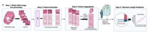
![The central thesis of TLPath is that telomere length can be determined from cell and tissue shape. Trained on more than 5,000 whole-slide images across 919 individuals and 18 organs, TLPath uses machine learning to detect architectural changes in tissues due to aging. These morphological features were used to accurately predict telomere length in 11 tissues and outperformed chronological age. [Anamika Yadav, Kyle Alvarez, Sanju Sinha, Sanford Burnham Prebys]](https://www.genengnews.com/wp-content/uploads/2026/03/Low-Res_TLPath-Cell-Press-Methods-300x72.jpeg)
Recent advances in computational pathology have demonstrated the potential to predict molecular properties, including mutation status, gene expression profiles, and chromosomal alteration, from high-resolution tissue images, the team continued. This points to the potential that evaluating cellular morphology from routinely available tissue histology might have predictive utility for determining tissue telomere length, without the need for specialized molecular techniques. “Considering that telomere length is a molecular hallmark of aging, the ability to quantify it from routine pathology images could significantly advance the field … We hypothesized that by systematically analyzing cellular morphology in wholeslide images, we could develop a model to predict bulk telomere length measurements.”
To develop TLPath, Sinha and team obtained data from the Genotype-Tissue Expression Project, a major National Institutes of Health (NIH) Common Fund initiative that launched in 2010 to create a resource for studying how inherited changes in genes lead to common diseases. The researchers trained their computational model on scans of 5,263 histopathology slides made from routine biopsy samples of 18 tissue types that were donated by 919 individuals.
“The dataset pairs these high-resolution images with laboratory tests of telomere length, enabling us to train TLPath to find predictive features in the cells and tissue,” explained Sinha. “There are hundreds of terabytes of imaging data from this project ripe for study with tools such as TLPath, and we could not have finished our project without this data being available to researchers.”
The model works by segmenting each histopathology slide into an average of 1,387 square fragments. Each fragment, known as a patch, is scoured to find up to 1,024 structural features. By computing a statistical weight for each feature on each patch, the model compares an overall score for each histopathology slide with the paired telomere length to learn how to predict the latter from the former.
After training TLPath separately on each tissue type, the scientists found it capable of predicting relative telomere length (RTL) on samples from the Genotype-Tissue Expression Project that had not been included in the training dataset. “Mechanistic interpretation of the model revealed that TLPath determines short telomere samples using senescence-like cell morphology (high nuclear-to-cytoplasmic ratio), along with tissue damage (necrosis) and fascia-like structures,” the authors stated.
“The key to our work was building on recent developments in computer vision for histopathology slides, which is the creation of foundation models,” said Sinha. “These models don’t look at discrete pixels, but instead define more higher-order features, only some of which can be interpreted by humans, yet can be validated for their predictive power … This opens up new opportunities based on the conceptual advancement that measurable structural changes in cells can predict the length of telomeres. Directly measuring telomere length requires more complicated and costly tests that are difficult to scale.”
While these slides are commonly created from biopsies for pathologists to review in the course of clinical care, they are rarely digitized and made available to researchers in a similar manner as the NIH-funded Genotype-Tissue Expression Project. “Whether it is new slides being developed today or those preserved in biobanks, all we need is for them to be properly scanned, stored, and shared in order to enable large-scale studies,” said Sinha.
To our knowledge, this is the first approach capable of predicting RTL from H&E slides, offering a new window into telomere research through machine learning analysis of tissue morphology,” the authors stated in their report. “TLPath outperformed chronological age in predicting telomere length, demonstrating H&E-based morphology’s ability to capture individual-specific telomere information.”
The post Computational Model Predicts Telomere Length from Images of Routine Histopathology Slides appeared first on GEN – Genetic Engineering and Biotechnology News.
GEN – Genetic Engineering & Biotechnology News
Bioengineered Implants Deliver Multi-Drug Therapy in Animal Models
In a new paper, scientists from Northwestern University and their collaborators at Rice University and Carnegie Mellon University report on their progress towards developing so-called implantable “living pharmacies.” These are tiny devices containing engineered cells that continuously produce medicines inside the body. Details of the study, which was done in rats, are published in Device in a paper titled “Design of a wireless, fully implantable platform for in-situ oxygenation of encapsulated cell therapies.”
The device, which is called the hybrid oxygenation bioelectronics system for implanted therapy or HOBIT, is roughly the size of a folded stick of gum. It integrates engineered cells with oxygen-producing bioelectronics and is designed in such a way that the cells are shielded from the body’s immune system while also receiving oxygen and nutrients needed to keep them alive and producing drugs for several weeks. In the future, these devices could be deployed to treat chronic conditions without requiring patients to carry, inject, or remember to take medications.
“This work highlights the broad potential of a fully integrated biohybrid platform for treating disease,” said Jonathan Rivnay, PhD, a professor of biomedical engineering and materials science and engineering at Northwestern and a co-principal investigator of the project. “Traditional biologic drugs often have very different half-lives, so maintaining stable levels of multiple therapies can be challenging. Because our implanted ‘cell factories’ continuously produce these biologics, keeping the cells alive with our oxygenation technology allows us to sustain steady levels [of] multiple different therapeutics at once.”
Solving the oxygenation challenge was critical to the success of HOBIT. When engineered cells are packed together in an implant, they compete for oxygen to live. Without a steady supply, many cells die, which limits how much medicine the implants can produce. In an earlier study, Rivnay and his collaborators demonstrated how a tiny electrochemical device could generate oxygen by splitting nearby water molecules, and showed that supplying oxygen locally dramatically improved the survival of implanted therapeutic cells. The latest iteration of their device integrates that oxygen-generation technology in a fully implantable, wireless system.
Digging into the details of the device, HOBIT contains three primary components: a cell chamber that holds the genetically engineered cells, a miniature oxygen generator, and electronics and a battery to regulate oxygen production and wirelessly communicate with external devices. Because the device produces oxygen directly inside the implant, the cells receive a steady supply even in hypoxic environments. “We are producing oxygen directly where the cells need it,” Rivnay said. “That allows us to support much higher cell densities in a much smaller space.” In fact, “cell densities in HOBIT were roughly six times higher than conventional unoxygenated encapsulation approaches.”
According to the paper, the team engineered the cells to produce three different biologics—an anti-HIV antibody, a GLP-1-like peptide used to treat type 2 diabetes, and leptin, a hormone that regulates appetite and metabolism. They implanted the devices under the skin of rats and monitored drug levels in their bloodstreams for 30 days. Blood measurements of animals with the implanted devices showed sustained levels of all three biologics throughout the study period. In contrast, in animals that were implanted with devices without oxygenation, the biologics that had shorter half-lives were undetectable by the seventh day. Drugs with longer half-lives in these animals also declined steadily over time. At the end of the testing period, roughly 65% of the cells in the oxygenated devices remained viable compared with roughly 20% in control devices.
For their next steps, the scientists intend to test their devices in larger animal models and explore disease-specific applications, including therapies based on transplanted pancreatic cells. “As these technologies continue to develop, devices like this could eventually act as programmable drug factories inside the body—delivering complex therapies in ways that simply aren’t possible today,” Rivnay said.
The post Bioengineered Implants Deliver Multi-Drug Therapy in Animal Models appeared first on GEN – Genetic Engineering and Biotechnology News.
GEN – Genetic Engineering & Biotechnology News
Gut-Immune Link Identified in Multiple Sclerosis-Related Neuroinflammation
Multiple sclerosis (MS) is a debilitating neurological disorder caused by malfunctioning immune responses that target the brain and spinal cord of the central nervous system (CNS). New research led by Shohei Suzuki, MD, PhD, assistant professor, division of gastroenterology and hepatology, and Tomohisa Sujino, PhD, associate professor, School of Medicine, at Keio University, Japan, has now indicated how the gut can initiate neuroinflammation in multiple sclerosis.
Their study found that intestinal epithelial cells (IECs) promote the development of pathogenic T cells that migrated to the spinal cord and induced disease symptoms in mouse models of the disorder.
The researchers examined intestinal tissues from patients with MS and mice with experimental autoimmune encephalomyelitis (EAE), a close analog of MS. In both cases, they observed an increase in TH17 cells and an upregulation of major histocompatibility complex class II (MHC II) expression in IECs. Deleting MHC II in IECs reduced the accumulation of TH17 cells in the gut and lowered the severity of EAE. They suggest the results could inform future strategies for developing targeted therapeutics against autoimmunity.
“While current therapies for MS often target B cells, our study highlights the gut as an important therapeutic site,” Suzuki commented. “Modulating intestinal microbiota or antigen-presenting activity of IECs represents new approaches to treating autoimmune neurological diseases.”
Suzuki, Sujino, and colleagues reported on their findings in Science Immunology, in a paper titled “Intestinal Epithelial MHC Class II Induces Encephalitogenic CD4⁺ T Cells and Initiates Central Nerves System Autoimmunity,” in which they concluded, “Our findings reveal an interaction between gut IECs and neuroinflammatory diseases through MHC II expression in human MS and mouse EAE, providing a mechanistic link between gut immune education and CNS autoimmunity and opening new avenues for targeting intestinal immunity in neuroinflammatory diseases.
Failure of the immune system to distinguish ‘self’ from ‘non-self’ entities leads to excessive autoimmune responses against self-proteins like myelin, which forms a protective covering on the neurons. Multiple factors influence the onset and progression of MS, including genetic susceptibility, environmental triggers, and, more recently, the gut microenvironment. Patients with MS exhibit alterations in their gut microbiota, while the gut microbiota and microbial metabolites play a pivotal role in shaping the chronic autoreactive immune responses. “… in an experimental autoimmune encephalomyelitis (EAE) model, commensal or specific microbes were found to be essential for disease initiation and progression,” the authors wrote.
However, in trying to define this gut–CNS axis, the cellular mechanisms that relay the gut-derived signals to the immune system to influence autoimmune inflammation in the CNS remain poorly understood. “Increasing evidence shows that the gut microbiota influences neurological diseases such as Parkinson’s, Alzheimer’s, and MS,” Sujino stated. “However, the mechanisms linking gut microbes, intestinal immunity, and brain inflammation remain unclear. We were keen to identify how gut immune responses contribute to neuroinflammatory diseases.”
Prior research has shown that gut-derived signals can promote the differentiation of T cells into pathogenic T helper 17 (TH17) in mouse models of MS. Recent studies have suggested that IECs can function as antigen presenting cells that help induce these pathogenic cells, but the underlying mechanisms have been unclear.
Building on their previous observation that mild intestinal (ileal) inflammation exists in experimental autoimmune encephalomyelitis (EAE), which is a mouse model of MS, the authors set out to test whether similar inflammation is present in patients with MS. By performing single-cell RNA sequencing on intestinal biopsies, the team identified that inflammatory Th17 cells accumulate in the mouse EAE model as well as in the intestine of patients with MS, suggesting a conserved gut–CNS axis that may be active in human diseases.
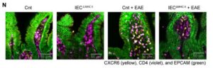
In both EAE mice and patients with MS, intestinal epithelial cells upregulated antigen presentation pathways. Particularly, epithelial cells in the ileum had higher expression of major histocompatibility complex class II (MHC II) that presents antigens to CD4+ T cells. “Clinically, patients with MS exhibited an increased expression of epithelial MHC II–associated genes and an accumulation of CD4 T cells in the small intestine, suggesting the conservation of this gut-CNS axis in human diseases,” the scientists stated. Experiments showed that selective deletion of MHC II in IECs reduced pathogenic Th17 cell generation and disease severity. “Conditional deletion of MHC II in IECs showed that epithelial antigen presentation was indispensable for the local expansion of pathogenic Th17 cells in the gut and their subsequent migration to the CNS,” the team stated.
![Immunofluorescence analysis was performed on terminal ileum samples from Cnt, IECΔMHCII, Cnt + EAE, and IECΔMHCII + EAE mice. A total of 3–5 tissue sections were analyzed per mouse, with 3 mice included in each group. [Shohei Suzuki]](https://www.genengnews.com/wp-content/uploads/2026/03/low-res-2-1-300x96.jpeg)
mouse, with three mice included in each group. [Shohei Suzuki]
IECs do not typically present antigens to immune cells. So, the team conducted co-culture assays to test the antigen presentation function of IECs. Their findings demonstrate that IECs can directly present antigens in an MHC II-dependent manner to prime CD4+ T cells in the gut. Notably, in these assays, IECs induced Th17 polarization of activated CD4+ T cells. It became clear that the gut was a critical site for immune activation of pathogenic CD4+ T cells that polarized into pro-inflammatory Th17 cells. “These findings provide direct functional evidence that IEC-expressed MHC II is sufficient to drive Th17 polarization from primed CD4 T cells in an antigen-dependent manner, supporting a direct role for IECs as non-professional antigen-presenting cells,” the scientists reported.
To investigate whether the Th17 cells directly contribute to the pool of autoreactive cells in the CNS, they used transgenic mice that express the Kaede protein, which undergoes photoconversion from green to red fluorescence upon exposure to violet light. This model allowed for precise tracking of pathogenic Th17 cells induced in the intestinal lamina propria that then migrate to the spinal cord and drive neuroinflammation.
Taken together, the study findings reveal a critical role for MHC II expressed by IECs in the expansion of pathogenic Th17 cells that subsequently migrate to the CNS during EAE, providing a mechanistic link between gut immune responses and autoimmune neuroinflammatory diseases. The results demonstrate that while systemic circulation allows T cell exchange across immune tissues, the epithelial–immune interactions within the gut mucosal compartment can essentially shape effector T cell responses in the brain.
“This study reveals a previously unknown role of IECs in antigen presentation and Th17 programming, thereby defining a gut-CNS immunological axis with important implications for understanding and treating autoimmune neuroinflammation,” the authors concluded. “Our findings suggest that the modulation of epithelial antigen presentation could serve as a novel therapeutic approach for MS and related diseases. Given the accessibility of the gut epithelium to dietary, microbial, and pharmacological interventions, targeting IEC–T cell interactions may offer a tractable strategy for immunomodulation.”
The post Gut-Immune Link Identified in Multiple Sclerosis-Related Neuroinflammation appeared first on GEN – Genetic Engineering and Biotechnology News.
GEN – Genetic Engineering & Biotechnology News
Agentic AI, Virtual Cell, LNP Vaccine Boosters, Engineered Organs, and Mergers
This week, agentic AI steps into the limelight buoyed by the momentum from generative AI. And there’s a new virtual cell model in town courtesy of AI-drug developer Xaira Therapeutics. From the frontiers of AI, our discussion turned to feats of engineering in regenerative medicine and lipid nanoparticles. In one study, scientists redesigned LNPs to avoid the liver and accumulate in the lymph nodes. In the other, efforts to develop and implant a lab grown esophagus from donor pigs bear fruit. Finally, Novartis plans to spend up to $3 billion to expand its cancer pipeline with the acquisition of Pikavation Therapeutics. And Merck is acquiring Terns Pharmaceuticals for approximately $6.7 billion also with an eye towards boosting its cancer portfolio.
Listed below are links to the GEN stories referenced in this episode of Touching Base:
NVIDIA GTC 2026: Agentic AI Inflection Hits Healthcare and Life Sciences
By Fay Lin, PhD, GEN Edge, March 18, 2026
Xaira’s First Virtual Cell Model Is Largest To-Date, Toward Complex Biology
By Fay Lin, PhD, GEN Edge, March 25, 2026
Modified Lipid Nanoparticles Boost mRNA Vaccine Delivery to Lymph Nodes
GEN, March 24, 2026
Engineered Esophagus Rebuilds Missing Organ Segment in Pig Models
GEN, March 20, 2026
Novartis Acquires Pikavation for Up to $3B, Expanding Cancer Pipeline
GEN, March 22, 2026
Merck Bolsters Cancer Pipeline with $6.7B Terns Buyout
By Alex Philippidis, GEN Edge, March 25, 2026
Touching Base Podcast
Hosted by Corinna Singleman, PhD
Behind the Breakthroughs
Hosted by Jonathan D. Grinstein, PhD
The post Agentic AI, Virtual Cell, LNP Vaccine Boosters, Engineered Organs, and Mergers appeared first on GEN – Genetic Engineering and Biotechnology News.


Illinois’ financial crisis could bring the state to a halt
The final 6 ‘Game of Thrones’ episodes might feel like a full season
New Season 8 Walking Dead trailer flashes forward in time
Mod turns ‘Counter-Strike’ into a ‘Tekken’ clone with fighting chickens
Meet Superman’s grandfather in new trailer for Krypton
Disney’s live-action Aladdin finally finds its stars

STAT+: FDA approves Sanofi diabetes drug for children with stage 3 diabetes
Opinion: ‘I’m pretty much all in’: An interview with a woman starting medical residency at almost 73

STAT+: Trump administration revisits policy to close Medicare drug price negotiation loophole
Lilly unveils first clinical data behind $2.3B Ajax deal, showing JAK inhibitor works ‘right out of the gate’
Hantavirus One-Shot mRNA Vaccine Fully Protects in Syrian Hamster Model
SonoThera Raises $125M to Develop Ultrasound-Mediated Genetic Medicines
Illinois’ financial crisis could bring the state to a halt
The final 6 ‘Game of Thrones’ episodes might feel like a full season
New Season 8 Walking Dead trailer flashes forward in time
Mod turns ‘Counter-Strike’ into a ‘Tekken’ clone with fighting chickens
Meet Superman’s grandfather in new trailer for Krypton
Disney’s live-action Aladdin finally finds its stars
-
 Uncategorized9 years ago
These ’90s fashion trends are making a comeback in 2017
-
Uncategorized9 years ago
According to Dior Couture, this taboo fashion accessory is back
-
Endpoints News3 months ago
Novartis to pay $2B upfront to take next-gen PI3Kα inhibitor from Synnovation
-
Uncategorized9 years ago
Phillies’ Aaron Altherr makes mind-boggling barehanded play
-
Uncategorized9 years ago
Uber and Lyft are finally available in all of New York State
-
Contributors9 years ago
The final 6 ‘Game of Thrones’ episodes might feel like a full season
-
Uncategorized9 years ago
Steph Curry finally got the contract he deserves from the Warriors
-
Uncategorized9 years ago
The old and New Edition cast comes together to perform