Uncategorized
Cancer Drug Shortfalls Tied to How BET Inhibitors Hit BRD2 and BRD4 Differently
For more than a decade, BET inhibitors have been touted as one of cancer therapy’s most promising drug classes. The logic was straightforward: many tumors rely on oncogenes that depend on BET (bromo- and extra-terminal domain) proteins—chromatin‑binding regulators that help switch genes on. Block the BET family, the thinking went, and cancer cells should lose their transcriptional fuel. In the lab, the strategy often worked. But in clinical trials, the results were far more uneven: modest responses, substantial side effects, and little clarity about which patients might benefit.
A new study from the Max Planck Institute of Immunobiology and Epigenetics (MPI‑IE) may finally explain why. Published in Nature Genetics, the work uncovers a previously underappreciated division of labor within the BET family—one that helps clarify why drugs that block all BET proteins at once have struggled in the clinic. The paper is titled, “Histone acetylation-dependent clustering of BRD2 instructs transcription dynamics.”
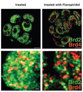
Most BET inhibitors were designed to block a shared bromodomain that all BET proteins use to bind chromatin. That approach assumed the proteins—BRD2, BRD3, BRD4, and BRDT—perform similar roles. But the new study paints a more nuanced picture. Using rapid protein degradation, chemogenomics, and super‑resolution microscopy in mouse embryonic stem cells, the team dissected the distinct contributions of BRD2 and BRD4 to transcription.

Their findings reveal that BRD4 drives the well‑known step of releasing paused RNA polymerase II into productive elongation. BRD2, however, acts earlier. It helps recruit and organize the transcription initiation machinery at promoters, particularly under conditions where pause‑release is impaired. As the authors wrote, BRD2’s role becomes “particularly critical under the conditions of impaired pause release,” a mechanistic insight that reframes how BET proteins collaborate during gene activation.
The MPI‑IE team likens BRD2 to a stage manager. “BRD2 sets up the stage: assembling the props, costumes, and actors to ensure preparations run smoothly. BRD2 then gives BRD4, the actor, the ‘start’ signal to begin with the performance,” said senior author Asifa Akhtar, PhD. Blocking both proteins simultaneously—exactly what current BET inhibitors do—disrupts two different steps of transcription at once, producing unpredictable and context‑dependent effects.
“Our data shows that the setup work happening before is just as critical for gene activation,” explained Akhtar.
A key discovery is that BRD2’s recruitment depends on histone H4 acetylation placed by the enzyme MOF. When MOF was rapidly depleted or deleted, BRD2 lost its grip on chromatin, while BRD3 and BRD4 remained largely unaffected. “The findings support a model in which acetylated chromatin creates a platform that allows regulatory proteins like BRD2 to concentrate and prepare the transcription machinery,” noted first author Umut Erdogdu, PhD.
The team also showed that BRD2 forms dynamic clusters at promoters. Removing only the BRD2 region responsible for clustering stalled transcription almost as completely as deleting the entire protein.
The study suggests a path forward: instead of blocking all BET proteins indiscriminately, future therapies may need to distinguish between BRD2‑ and BRD4‑specific functions. “Thus, these findings support a model in which histone acetylation-dependent spatiotemporal dynamics of BRD2 coordinate the transcription machinery to regulate transcription initiation,” the authors wrote.
For a field long puzzled by the uneven performance of BET inhibitors, BRD2’s newly revealed role offers a compelling piece of the puzzle—and a clearer blueprint for next‑generation cancer therapeutics.
The post Cancer Drug Shortfalls Tied to How BET Inhibitors Hit BRD2 and BRD4 Differently appeared first on GEN – Genetic Engineering and Biotechnology News.
Uncategorized
Amgen shores up Tavneos’ FDA defense with Duke data analysis
Amgen shores up Tavneos’ FDA defense with Duke data analysis
After the FDA flagged patient deaths linked to Amgen’s rare disease drug Tavneos and called for its voluntary removal, the pharma recruited an independent data analysis from Duke researchers to help build the case for the drug’s continued market approval. Read More
Uncategorized
Chile offers new data on food warning label efficacy
Get your daily dose of health and medicine every weekday with STAT’s free newsletter Morning Rounds. Sign up here.
So much news today that I didn’t have space to write an item about hot tubs as a breeding ground for Legionnaires’ disease. Here’s the CDC report, if you’re curious.

Get your daily dose of health and medicine every weekday with STAT’s free newsletter Morning Rounds. Sign up here.
So much news today that I didn’t have space to write an item about hot tubs as a breeding ground for Legionnaires’ disease. Here’s the CDC report, if you’re curious.
The RNA-based medicine is one of a handful of antibody-oligonucleotide conjugates that Novartis acquired last October when it took over neuromuscular-focused Avidity Biosciences.


Illinois’ financial crisis could bring the state to a halt
The final 6 ‘Game of Thrones’ episodes might feel like a full season
New Season 8 Walking Dead trailer flashes forward in time
Mod turns ‘Counter-Strike’ into a ‘Tekken’ clone with fighting chickens
Meet Superman’s grandfather in new trailer for Krypton
Disney’s live-action Aladdin finally finds its stars
Amgen shores up Tavneos’ FDA defense with Duke data analysis

Chile offers new data on food warning label efficacy
Novartis’ $12B Avidity buy pays dividends with Phase 1/2 muscular dystrophy win

STAT+: At hospital finance conference, a call to end the friction that’s keeping costs high
Beyond sunshine: Iberia’s biotech moment has arrived with developing capital networks

Laser‑Driven Phase Contrast Enhances Cryo‑EM Resolution of Small Proteins
Illinois’ financial crisis could bring the state to a halt
The final 6 ‘Game of Thrones’ episodes might feel like a full season
New Season 8 Walking Dead trailer flashes forward in time
Mod turns ‘Counter-Strike’ into a ‘Tekken’ clone with fighting chickens
Meet Superman’s grandfather in new trailer for Krypton
Disney’s live-action Aladdin finally finds its stars
-
 Uncategorized9 years ago
These ’90s fashion trends are making a comeback in 2017
-
Uncategorized9 years ago
According to Dior Couture, this taboo fashion accessory is back
-
Endpoints News3 months ago
Novartis to pay $2B upfront to take next-gen PI3Kα inhibitor from Synnovation
-
Uncategorized9 years ago
Phillies’ Aaron Altherr makes mind-boggling barehanded play
-
Uncategorized9 years ago
Uber and Lyft are finally available in all of New York State
-
Contributors9 years ago
The final 6 ‘Game of Thrones’ episodes might feel like a full season
-
Uncategorized9 years ago
Steph Curry finally got the contract he deserves from the Warriors
-
Uncategorized9 years ago
The old and New Edition cast comes together to perform