Uncategorized
Brain Astrocytes Form Far-Reaching Connections in Mice
A study in mice headed by NYU Langone Health researchers has found that cells long thought to play a secondary role in brain function build their own far-reaching connections. These pathways appear to connect distant regions in ways that had not been mapped before.
Experts usually describe the brain as a network of nerve cells (neurons) that send each other signals to pass along information. These neurons are maintained by another kind of brain cell, the star-shaped astrocyte, which ferries in nutrients and carries away waste.
The newly reported study, headed by Melissa Cooper, PhD, a postdoctoral fellow in the department of neuroscience at NYU Grossman School of Medicine, revealed that, like neurons, astrocytes form organized webs, which enable them to communicate with other specific astrocytes across the brain rather than only sending local, generalized signals. In some cases, the pathways were found to link areas that were not already joined together by neurons.
“For more than a century, neuroscientists have thought of neurons as the main actors in the brain,” said Cooper. “Yet our findings suggest that astrocytes, which are usually viewed as merely support cells, are also running their own widespread signaling pathway, adding another layer to how brain regions stay connected.” The team suggests that while their study was carried out in mice, not humans, the findings form the basis for future studies investigating how astrocyte networks might link with injury, disease, or aging and to learning and memory.”
Cooper is first and co-corresponding author of the team’s published work in Nature, titled “Astrocytes connect specific brain regions through plastic networks,” in which the researchers stated, “Astrocyte networks can directly link brain regions that are not connected by neurons, suggesting that previously unassociated brain regions communicate with one another through gap junction-coupled astrocytes.”
“Neuronal axons have traditionally been considered to be the primary mediators of functional connectivity among brain regions,” the authors wrote, and the role of communication mediated by astrocytes has been largely underappreciated. “This communication occurs through gap junctions—membrane channels that connect the cytoplasm of neighboring cells, enabling them to redistribute resources and share biochemical signals,” the team continued. “Studies using mice lacking astrocyte gap junctions have shown that these gap junctions are necessary for memory formation, synaptic plasticity, coordination of neuronal signaling, and closing the visual and motor critical periods.”
In earlier work, Cooper reported that in a mouse model of the visual neurodegenerative disease glaucoma, astrocytes can redistribute resources from astrocytes around healthy neurons to damaged neurons. Yet the team had no way to see whether this kind of support-cell network extended across the entire brain.
Cooper said the newly reported study is the first to map active, brain-wide communication networks built by astrocytes and to show that these pathways are highly specific. The research relied on a custom-built tracing tool that let the team follow the cells’ connections in far greater detail than had been possible using past methods. “Despite the importance of astrocyte gap junctional networks, studying them has been challenging,” the investigators noted. “Current methods such as slice electrophysiology disrupt network connectivity and introduce artefacts due to tissue damage.”
For their study, the researchers used a harmless virus to deliver “network tracers” into astrocytes in selected brain regions of lab mice. These tracers tagged small molecules as the molecules passed through the gap junctions linking one astrocyte to another, allowing the team to see which cells were part of the same signaling pathway.
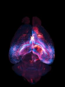
The scientists then made the mice’s brains transparent and used a specialized microscope to capture three-dimensional images of every tagged astrocyte. By doing this across hundreds of mice, they could map astrocyte webs across brain areas. “These networks selectively connect specific regions, rather than diffusing indiscriminately, and vary in size and organization,” they reported. “We observe local networks that are confined to single brain regions and long-range networks that robustly interconnect multiple regions across hemispheres, often exhibiting patterns distinct from known neuronal networks.”
![A 3D network of interconnected astrocytes imaged inside a whole, transparent mouse brain. Each astrocyte's color shows its distance from the viewer; closer astrocytes are blue, while more distant astrocytes are red. [Cooper et al. Astrocytes connect specific brain regions through plastic networks. Nature. 2026. doi:10.1038/s41586-026-10426-6.]](https://www.genengnews.com/wp-content/uploads/2026/04/Low-Res_Cover-Image-224x300.jpg)
The tracing tool and brain-clearing method were designed to be relatively low-cost and easy to reproduce so that other labs could use them to study the networks in many brain diseases.
In another part of the study, the team assessed mice that were genetically engineered with astrocytes that lacked gap junctions. The communication networks largely disappeared, suggesting that the pathways are active and depend on these physical bridges.
“By challenging our understanding of how the brain communicates over long distances, our results may offer fresh insight into how it develops, ages, and behaves in conditions such as Alzheimer’s and Parkinson’s diseases,” said study co-senior author Shane A. Liddelow, PhD, an associate professor in the neuroscience and ophthalmology departments at NYU Grossman School of Medicine.
Another key finding was that astrocyte networks are dynamic. When the team trimmed whiskers on one side of the mice’s faces—“this manipulation is known to induce robust structural remodeling in neurons,” the team noted—a pathway from the region that processes whisker touch got smaller and reconnected to different astrocyte partners.
“The fact that astrocyte networks shrink and reroute after a loss of sensory signals suggests they may be shaped by experience,” said study co-senior author Moses V. Chao, PhD, a professor in the cell biology, neuroscience, and psychiatry departments at NYU Grossman School of Medicine. “It also raises the possibility that each of us has a somewhat unique pattern of connections molded by what our brains have learned and lived through.”
The authors plan to investigate which molecules move through the networks and to apply their tracing tool to models of brain disorders. They also hope to examine how these webs change during development and aging, said Chao.
Liddelow emphasized that while gap junctions and astrocytes exist in humans, it remains unknown whether the networks link the same regions in the same way as in mice. Nevertheless, in their paper, the team concluded that their findings “… establish foundation for future exploration of how astrocyte network structure and function are shaped by injury, disease, development, aging and experience-dependent processes such as learning and memory.”
The post Brain Astrocytes Form Far-Reaching Connections in Mice appeared first on GEN – Genetic Engineering and Biotechnology News.
Uncategorized
STAT+: Trump administration revisits policy to close Medicare drug price negotiation loophole

WASHINGTON — The Trump administration on Friday proposed to change a policy that is designed to prevent drugmakers from avoiding Medicare price negotiation by adding active ingredients to drugs.
The policy is part of an annual proposed rule that establishes the process that the Centers for Medicare and Medicaid Services uses to choose the next 20 drugs and biologics for price negotiation. Those drugs will be announced by Feb. 1, 2027, and their negotiated prices will take effect in 2029. The administration also considered a similar policy last year but put off a decision to study it further.
Medicare must wait seven to 11 years after a product is approved by the Food and Drug Administration before it can negotiate its price, depending on the type of medicine. Biologics that are typically administered in doctor offices get more time than drugs taken orally.
Last month, the Andes virus outbreak on a Dutch cruise ship departing from Argentina brought a transmission context for hantavirus, that was previously unprecedented, to the forefront. The Andes virus is the only member of the hantavirus family that is capable of efficient person-to-person spread through close contact with respiratory secretions. Other hantaviruses are typically spread through contact with infected rodents, making the Andes virus a much more significant public health threat.
While at sea, the outbreak spread among passengers and crew, infecting 13 people and killing three. The cruise passengers have since returned to their home countries, 23 in total. Because a person can carry the virus for weeks before showing any symptoms, health agencies are facing a complex challenge of identifying everyone who was exposed. There are currently no vaccines or preventive treatments approved for the virus; this travel-related outbreak brought the need for vaccine development to the forefront.
Researchers at The University of Texas Medical Branch (UTMB) had previously developed and tested two mRNA vaccines against intramuscular Andes virus challenge in golden Syrian hamsters (“1-methylpseudouridine-modified or non-modified mRNA modalities encoding the envelope glycoproteins, Gn and Gc, in a single open reading frame.”)
When tested in the Syrian hamster model, both mRNA vaccines were efficacious in hamsters using a two-dose regimen. Recognizing that a fast-moving international outbreak doesn’t allow time for patients to wait weeks between shots, the team retested the vaccines to determine whether a single dose would be effective.
Now, a new report shares the finding that the vaccine provided full protection against the Andes hantavirus after a single dose.
This work is published in The Lancet in the paper, “Single-dose mRNA vaccines against Andes hantavirus.”
Alexander Bukreyev, PhD, head of the Laboratory of Viral Pathogenesis and Vaccine Development at UTMB, said that the group is working to fast-track these single-dose vaccines into human clinical trials.
The results exceeded expectations. When testing the vaccines in an animal model that mimics human disease, the scientists found that a single shot provided 100% protection against a lethal dose of the virus. Even when the researchers significantly lowered the dosage to a fraction of the original amount, the results remained definitive.
“Every vaccinated animal remained completely healthy and showed no symptoms or weight loss,” said Michelle Meyer, PhD, senior scientist in the Bukreyev Laboratory. “When we looked at the tissues from the vaccinated animals a month after infection, the virus was entirely gone. The vaccines triggered a powerful immune response, creating protective antibodies in as little as 14 days.”
Because the Andes virus can take a relatively long time to make a human severely ill, these fast-acting vaccines could serve a dual purpose, possibly functioning as an emergency tool for people who have already been exposed.
“If given quickly to high-risk contacts during an outbreak, such as the Andes virus situation on the cruise ship, the vaccines could theoretically jump-start their immune systems fast enough to intercept the virus—stopping it from replicating and preventing them from getting sick or spreading it further,” Bukreyev said.
The post Hantavirus One-Shot mRNA Vaccine Fully Protects in Syrian Hamster Model appeared first on GEN – Genetic Engineering and Biotechnology News.
Biotechnology company SonoThera has raised $125 million in an oversubscribed Series B financing round. The financing was led by Vida Ventures, with participation from ARK Invest, CureDuchenne Ventures, Leaps by Bayer, Otsuka Pharmaceutical, SymBiosis, UCB Ventures SA, Vivo Capital, and existing investors ARCH Venture Partners, Alexandria Venture Investments, Duquesne Family Office, Illumina Ventures, Johnson & Johnson Innovation – JJDC, Medical Excellence Capital, RA Capital, and Vertex Ventures HC.
SonoThera will use the funds to advance its lead programs in Duchenne muscular dystrophy (DMD) and autosomal dominant polycystic kidney disease (ADPKD) in the clinic. The funds will also support efforts to expand its pipeline of targeted redosable genetic medicines across multiple organ systems and scale its proprietary platform technologies for safe, targeted therapy delivery.
The company’s platform combines a proprietary ultrasound-mediated delivery technology dubbed RIPPLE , with a payload engineering platform dubbed PORE. The platforms are designed to support the development of DNA and RNA therapeutics, gene editing, and gene silencing approaches. SonoThera is using its tech to develop genetic medicines that it claims will address key limitations of conventional gene therapies including delivery challenges, payload size constraints, immune responses, safety events, and difficulties with redosing.
As Kenneth Greenberd, PhD, SonoThera’s co-founder and CEO, stated “we founded SonoThera to take a fundamentally different approach, with a platform designed to broaden the therapeutic possibilities of the field. We believe our technology has the potential to expand the range of diseases addressable by genetic medicines while enabling more precise, durable, safer, and repeatable therapies for patients.”
SonoThera has already demonstrated the targeted delivery and expression capabilities of its platform across multiple tissues, including skeletal muscle, heart, liver, kidney, adipose, and brain. It has also shown that it can deliver large payloads such as full-length dystrophin for DMD and RNA-based payloads for gene silencing applications in preclinical studies.
The company expects to initiate its first clinical trial in DMD in 2027.
Commenting on the financing, Rajul Jain, MD, managing director at Vida Ventures, said “we believe SonoThera, with its RIPPLE delivery and PORE payload engineering technologies, has the potential to unlock opportunities in diseases with significant unmet need that have been previously inaccessible to other genetic medicine approaches.”
In connection with the financing, Jain and Rakhshita Dhar, MS, vice president & head of Healthcare Venture Investments at Leaps by Bayer, have joined SonoThera’s Board of Directors.
The post SonoThera Raises $125M to Develop Ultrasound-Mediated Genetic Medicines appeared first on GEN – Genetic Engineering and Biotechnology News.


Illinois’ financial crisis could bring the state to a halt
The final 6 ‘Game of Thrones’ episodes might feel like a full season
New Season 8 Walking Dead trailer flashes forward in time
Mod turns ‘Counter-Strike’ into a ‘Tekken’ clone with fighting chickens
Meet Superman’s grandfather in new trailer for Krypton
Disney’s live-action Aladdin finally finds its stars

STAT+: Trump administration revisits policy to close Medicare drug price negotiation loophole
Hantavirus One-Shot mRNA Vaccine Fully Protects in Syrian Hamster Model
SonoThera Raises $125M to Develop Ultrasound-Mediated Genetic Medicines

STAT+: Up and down the ladder: The latest comings and goings
FDA imposes import alert on Indian plant after inspectors flag GMP failings
Rapport ‘fully prepared’ to launch seizure drug solo, CEO says
Illinois’ financial crisis could bring the state to a halt
The final 6 ‘Game of Thrones’ episodes might feel like a full season
New Season 8 Walking Dead trailer flashes forward in time
Mod turns ‘Counter-Strike’ into a ‘Tekken’ clone with fighting chickens
Meet Superman’s grandfather in new trailer for Krypton
Disney’s live-action Aladdin finally finds its stars
-
 Uncategorized9 years ago
These ’90s fashion trends are making a comeback in 2017
-
Uncategorized9 years ago
According to Dior Couture, this taboo fashion accessory is back
-
Endpoints News3 months ago
Novartis to pay $2B upfront to take next-gen PI3Kα inhibitor from Synnovation
-
Uncategorized9 years ago
Phillies’ Aaron Altherr makes mind-boggling barehanded play
-
Uncategorized9 years ago
Uber and Lyft are finally available in all of New York State
-
Contributors9 years ago
The final 6 ‘Game of Thrones’ episodes might feel like a full season
-
Uncategorized9 years ago
Steph Curry finally got the contract he deserves from the Warriors
-
Uncategorized9 years ago
The old and New Edition cast comes together to perform