

Uncategorized
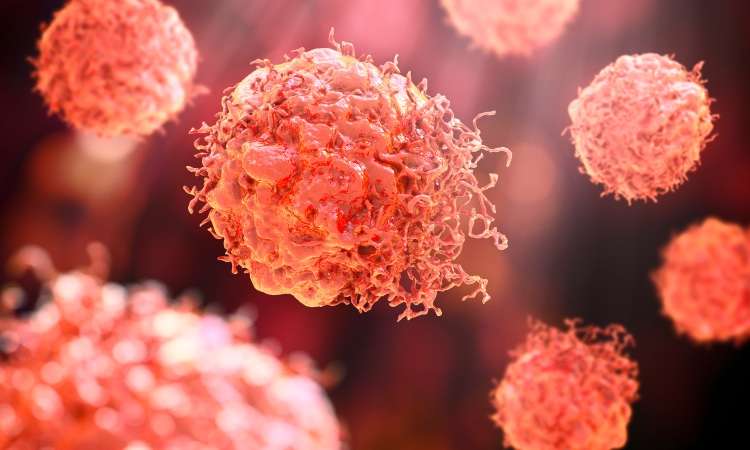
Human Multi-Organ Chip Offers New Insight Into Cancer Metastasis

Cancer metastasis is responsible for at least two-thirds of cancer deaths. Drugs targeting the metastatic progression have largely failed, in part due to the lack of predictive models that would help identify the underlying mechanisms of metastasis.
Now, new work reports the development of a multi-organ chip that mimics how cancer cells spread from vascular flow to distant organs—the first model of cancer metastasis of its kind. The chip includes compartments with millimeter-sized engineered human bone and lung tissues, and the vascular flow that contains circulating breast cancer cells and allows the dynamic cross-talk of the tissues being colonized.
“The key advantages of this advanced model of metastasis are that it is human and can be patient-specific,” said Gordana Vunjak-Novakovic, PhD, university professor and professor of biomedical engineering and professor of medical sciences at Columbia University. “It faithfully mimics some of the key aspects of human metastasis that are otherwise largely inaccessible for direct study.”
This work is published in Science Translational Medicine in the paper, “Organ-specific colonization and niche remodeling in a human tissue model of metastasis.”
“The pressing need for developing human tissue models of metastasis has been a key motivation for our study,” said Vunjak-Novakovic. “Our objective was to probe the ability of cancer cells to adhere to and traverse across endothelium [inner lining of blood vessels], and to determine their capacity to survive in the tissues they are colonizing through cell reprogramming and niche remodeling.”
The study sheds light on a critical phase of metastasis, known as organ colonization, which is difficult to study using animal models. The process is highly complex, requiring the cancer cells to evade tissue defense and adapt to the specific organs they invade.
The multi-organ chip allows scientists to investigate, in detail, metastatic progression with patient cells and tissues. The platform enables controlled experimentation of cancer cell-tissue interactions within organ-specific microenvironments, toward revealing molecular pathways and therapeutic targets for metastasis.
To demonstrate the chip’s capabilities, the team examined the colonization of circulating human breast cancer cells in bone and lung. Bone and lung, common sites of metastasis for breast cancer, and the vascular endothelium were engineered from induced pluripotent stem cells (iPSCs) using tissue-specific scaffold-bioreactor culture systems. The engineered tissues were maintained in individual compartments of the chip, which were each optimized for tissue maturation and long-term maintenance of functionality, and linked to each other by vascular circulation.
A selectively permeable endothelial barrier separates tissue compartments from the vascular channel, as in the human body. Once the platform was established, the researchers introduced breast cancer cells into the vascular circulation to observe patterns of organ-specific colonization. In line with what happens inside the human body, cancer cells that typically gravitate toward the bone showed stronger bone colonization and induced more pronounced bone degeneration. In contrast, cancer cells that typically gravitate toward the lung caused greater disruption in the lung tissue and only modest colonization of bone. Distinct patterns of tissue colonization and secreted factors demonstrate that this device mimics key features of organ-specific metastasis observed in the human body.
In addition, a post-analysis of the engineered tissue revealed that cancer cells condition the distant organs to be more receptive to colonization. The team saw signs of this process—called pre-metastatic niche formation—across both tissue compartments.
“Cancer is very smart, unfortunately. We learned how the cells cross barriers to get from blood circulation into the tissues,” said Vunjak-Novakovic. “We were also able to reproduce something that happens in patients, where cancer cells condition the target tissues, even before they colonize them, to make them more receptive.”
The work is aligned with the trend of more preclinical research in the future, including engineered human tissues that can complement what is learned from animal models. “As the FDA and NIH place growing emphasis on new approach methodologies, this study is a concrete example of what that shift can look like in practice, applied to one of cancer’s most challenging hallmarks: metastasis,” noted Ilaria Baldassarri, a PhD student at Columbia University.
The post Human Multi-Organ Chip Offers New Insight Into Cancer Metastasis appeared first on GEN – Genetic Engineering and Biotechnology News.


Hosted by Drug Discovery World and supported by Sartorius and BioIVT, this webinar will explore the opportunities and challenges that exist within cancer research drug discovery and development.
You will hear from Dr Sudha Rao, Chief Scientific Officer of Kazia Therapeutics, Karol Budzik, PhD, Business Development Associate at Vyriad Therapeutics and Lars van der Veen, Chief Scientific Officer at iOnctura.
Presentations will cover how cancer treatments have shifted towards reprogramming the biology driving tumour growth, immune escape and treatment resistance, the trajectory that in vivo CAR-T treatments are taking, and how challenging tumours burdened by stroma and immune-mediated resistance can be tackled.
Q&A with the speakers follows the presentations.
The post New webinar: Tackling drug discovery challenges in cancer research appeared first on Drug Discovery World (DDW).
Amid the rise of psychedelics in the mental health space, researchers have begun to explore psilocybin as a treatment for chemotherapy-induced peripheral neuropathy.
Uncategorized
New Spectrometry Technique Could Aid Formulation Development
New Spectrometry Technique Could Aid Formulation Development
A new technique combining two forms of spectrometry could help biopharmaceutical companies improve their choice of formulation buffer for antibody manufacturing by revealing how molecular forms and three-dimensional shapes of complex biologics respond to their environment. That’s the view of Christian Bleiholder, PhD, a professor at Florida State University who helped develop the technique.
According to Bleiholder, what happens structurally when a complex biological molecule, such as an antibody or viral spike protein, binds to its target is currently poorly understood.
“This is where [this approach] can help with the bioprocessing and formulation,” he says, as structural changes “can affect the lifespan [of the product] and lead to issues, such as aggregation.”
Because antibodies are complex, existing techniques tend to be powerful at different levels of complexity, he explains. Mass spectrometry is particularly powerful for distinguishing molecular composition, while structural approaches such as X-ray crystallography and cryo-electron microscopy can provide high-resolution structural information.
The challenge is understanding the link between these things within a heterogeneous sample, he says.
To overcome this, Bleiholder and his team worked with Bruker Daltonics to develop Tandem-Trapped Ion Mobility Spectrometry (Tandem-TIMS). This combines tandem ion mobility spectrometry with tandem mass spectrometry to disentangle three overlapping layers of molecular complexity: molecular form, three-dimensional shape, and binding or assembly state, he says.
He explains that, if the proteins have different structures, they can be characterized with tandem ion mobility spectrometry, and then mass spectrometry can be used to look at their molecular forms and binding states.
Going forward, Bleiholder hopes the technique can be used for formulation development but also earlier, during drug discovery of new products, such as multi-specific antibodies, to determine which molecular states are important and how those change when a biologic engages its target. He also plans to look at automating the technique.
Bleiholder spoke about using Tandem-TIMS at the Bioprocessing Summit in Boston earlier this year.
The post New Spectrometry Technique Could Aid Formulation Development appeared first on GEN – Genetic Engineering and Biotechnology News.


Single Hyaluronic Acid Injection Matches Three-Dose Therapy for Knee Osteoarthritis

Blood and Gut Cell Analyses Match 140 Inflammatory Bowel Disease Risk Loci

Study Details Chlamydia and Gonorrhea Co-Infection Rates in Hospital Patients

Ribociclib shows promise for specific recurrent endometrial cancer subtypes in clinical trial

FDA again turns back Capricor Duchenne cell therapy

Beat AML Precision Trial Marks Decade of Genomic Matching and Extended Survival
-

 Uncategorized8 months ago
Novartis buys Synnovation’s PI3Kα inhibitors for $3 billion
-
Endpoints News8 months ago
Novartis to pay $2B upfront to take next-gen PI3Kα inhibitor from Synnovation
-

 Uncategorized7 months ago
Optical Pooled CRISPR Screen Reveals Regulators of NF-κB Dynamics in Human Cells
-
Uncategorized6 months ago
Relay’s PI3Kα inhibitor clears efficacy bar in Phase 2 vascular anomalies study
-
Uncategorized6 months ago
Hybrid solid−liquid optics enable scalable, high-resolution light-sheet microscopy across diverse immersion media
-
Fierce Biotech8 months ago
Scientists turn pig semen extract into eye drops that kill cancer in mice
-
Nature Biotechnology8 months ago
Sustained nitric oxide production by engineered E. coli remodels the tumor microenvironment and potentiates immunotherapy
-
Nature Biotechnology8 months ago
Mapping cis-regulatory mutations at scale in sorghum enables modulation of gene expression
 Scan to Read