Uncategorized
Common Ancestry Limits Protein Sequence Exploration, Computational Study Shows
For all the excitement surrounding AlphaFold and the surge of AI‑driven protein design tools, one fact often goes unexamined: nearly every model is trained on databases of known proteins. These databases feel vast, but compared to the astronomical number of sequences that could, in principle, form a functional protein, they represent only a sliver of what’s possible. That raises a fundamental question for the field: how representative is the protein universe we currently know?
A new study published in PNAS and titled “Descent from a common ancestor restricts exploration of protein sequence space,” takes direct aim at that question, using large‑scale protein evolution modeling to probe the limits of protein diversification. The work, led by researchers at the Okinawa Institute of Science and Technology (OIST), the Institute of Science and Technology Austria (ISTA), the University of Vienna, and the Centro de Astrobiología (CAB), suggests that the boundaries of today’s protein diversity were set remarkably early in life’s history.
“Modern AI methods are thought to be revolutionizing protein design,” said Fyodor Kondrashov, PhD, who heads OIST’s Evolutionary and Synthetic Biology Unit, in a press release. “Yet most of these AI design methods are typically trained on databases of known proteins. Without understanding how representative these known proteins are of sequence space, how confident can we be that such methods can generate truly diverse protein designs?”

To investigate that representativeness, the team began by mathematically describing the region of sequence space occupied by known proteins. They estimated the “dimensionality” of each protein family, a measure of how many independent directions evolution has actually sampled, using correlation‑based analyses of sequence variation. The researchers then simulated protein evolution to test how ancestry, selection, and epistasis shape the diversity of sequences that evolution can reach. The goal was to estimate how many functional sequences should exist for a given protein family and to compare that theoretical diversity with the diversity observed in nature.
What emerged was a striking pattern: the strongest constraint on protein evolution is not selection or epistasis, but ancestry itself. Proteins tend to remain clustered near the sequences of their earliest ancestors, with limited divergence into the broader functional landscape. As the authors report, “For some gene families the effective topological dimension was on the order of one; in just a few families it was larger than 10.”
“That [the] starting point is the main evolutionary limit is not necessarily surprising, but the scale of its importance is really quite remarkable,” said lead author Lada Isakova, a PhD student within the unit. “As an evolutionary biologist, I was intrigued to see how little selection and epistasis seemed to matter in our results.”
The findings also feed into long‑standing debates about how the first proteins emerged. The team’s simulations suggest that early protein families could not have arisen simply by mutating a single first sequence. Instead, the data point toward DNA recombination “to create new DNA molecules which could encode very different proteins,” explained Isakova.
For today’s protein engineers, the work underscores a practical limitation: AI models trained on existing proteins may struggle to extrapolate into unexplored regions of sequence space. “Most methods won’t be able to generalize well beyond the current known sequence space,” Isakova noted. “We can see there are huge swaths of sequence space left to be explored, but it’ll take new experimental data to enable expansion into these unknown realms.”
The post Common Ancestry Limits Protein Sequence Exploration, Computational Study Shows appeared first on GEN – Genetic Engineering and Biotechnology News.

You know when you are at the eye doctor getting an updated prescription, and suddenly the world snaps into sharper focus? Physicists at the University of California (UC), Berkeley, have now done something similar for electron microscopy. By introducing phase contrast into a cryo‑electron microscope, they have delivered dramatically sharper images of some of biology’s smallest and most elusive proteins.
The advance comes from a new laser phase plate (LPP), described in the paper “Laser phase plate improves structure determination of small proteins by cryo‑EM,” which was published recently in Science. Led by physicist Holger Mueller, PhD, of UC Berkeley and Lawrence Berkeley National Laboratory, the team demonstrated that a laser‑driven phase plate can overcome one of cryo‑EM’s most persistent limitations: poor contrast for small proteins.

Cryo‑EM has transformed structural biology over the past decade, earning a Nobel Prize in 2017 for enabling high‑resolution structures without crystallization. But despite its impact, the technique still struggles with proteins below ~70 kilodaltons—a size range that includes about 90% of the human proteome. “Because of signal-to-noise limitations, the majority of human and animal proteins are too small to be analyzed by these methods [cryo-EM and cryoelectron tomography]. The increase in signal-to-noise ratio provided by this laser phase plate is expected to overcome these important limitations.”
The new LPP begins to address that problem. The LPP uses an intense, continuous‑wave laser to shift the phase of the electron beam itself. This produces true phase contrast without dimming or destabilizing the beam. Mueller described the laser focus as “75 kilowatts focused to a few microns… That’s more powerful than what you use for welding. It has more power than a military laser. It builds up the brightest continuous laser focus ever.”
Installed in a custom Thermo Fisher Titan Krios, the LPP immediately improved the clarity and resolvability of small proteins, including hemoglobin, which sits at the lower limit of what today’s cryo‑EM instruments can handle. As the authors wrote in the abstract: “Here, we show that the laser phase plate (LPP)… enhances the resolution in single-particle reconstruction of small proteins by improving specimen-motion correction, recovery of information from the early frames, as well as particle visualization, 3D classification, and alignment.”

These improvements were achieved using standard defocus ranges and reconstruction workflows. “For the most challenging cases—small particles, bad specimens—the laser produces a very considerable advantage,” Mueller said.
The impact extends beyond single‑particle analysis. Cryo‑electron tomography (cryo‑ET), which assembles multiple angular views of a molecule or protein into a three-dimensional image, stands to benefit even more. “With cryo-ET, we’re looking at small, very complicated cellular material that’s incredibly crowded inside the cell,” said Bridget Carragher, PhD, founding technical director of imaging at Biohub. “It’s like a forest of trees, and you’re trying to find one leaf on one tree in there. Cryo-ET needs a dramatic step forward in contrast, so we can start to see what’s going on inside the cell. That’s what the laser phase plate promises to give us.”
Biohub is developing a dual‑laser version of the system, designed to reduce component wear and minimize aberrations. Meanwhile, Mueller’s team is pushing toward imaging proteins as small as 17 kilodaltons, a threshold that would open access to vast regions of the human proteome previously invisible to cryo‑EM.
“This technology is a step function change for biology,” said Stephani Otte, PhD, Biohub’s vice president of imaging science. “What was once invisible will become visible—and that changes everything about how we understand disease.”
“The bottom line is, if you have a large protein and a really good sample—a fresh one or one frozen without bubbles, for example—you may not need the phase plate to get a single, high-quality image. But for a small protein and a bad sample, laser-on is best,” Mueller said. “This could fill an enormous gap in our knowledge of protein structures that can’t be crystallized or are too small for today’s cryo-EM. And it will be revolutionary for cryo-ET.”
The post Laser‑Driven Phase Contrast Enhances Cryo‑EM Resolution of Small Proteins appeared first on GEN – Genetic Engineering and Biotechnology News.
Uncategorized
STAT+: Updated: Tracking RFK Jr.’s promises to remake health in America
Updated June 11, 2026
WASHINGTON — A pledge to “Make America Healthy Again” earned Robert F. Kennedy Jr. his job atop U.S. health agencies a year and some change ago. He’s now had the opportunity to turn his words into action, with mixed results.
“All one needs” to prove the health secretary’s attentiveness is to “review my unprecedented list of accomplishments on a wide range of issues, all of which I drove,” Kennedy posted on X on Wednesday in response to a journalist.

Can any of the new obesity medications in development stand out from the pack? Which company just broke records with its IPO? And will the Food and Drug Administration allow greater access to experimental peptides?
We discuss all that and more on this week’s episode of “The Readout LOUD,” STAT’s biotech podcast.


Illinois’ financial crisis could bring the state to a halt
The final 6 ‘Game of Thrones’ episodes might feel like a full season
New Season 8 Walking Dead trailer flashes forward in time
Mod turns ‘Counter-Strike’ into a ‘Tekken’ clone with fighting chickens
Meet Superman’s grandfather in new trailer for Krypton
Disney’s live-action Aladdin finally finds its stars

Laser‑Driven Phase Contrast Enhances Cryo‑EM Resolution of Small Proteins

STAT+: Updated: Tracking RFK Jr.’s promises to remake health in America

An obesity drug deep-dive, and peptides move mainstream

RFK Jr. claims his calendar is publicly available. We’ve been trying to get it for a year

Nonprofit buys experimental cancer drug to maintain patient access
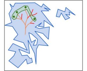
Potential Cocaine Addiction Targets Identified Through Genetic Mapping in Rats
Illinois’ financial crisis could bring the state to a halt
The final 6 ‘Game of Thrones’ episodes might feel like a full season
New Season 8 Walking Dead trailer flashes forward in time
Mod turns ‘Counter-Strike’ into a ‘Tekken’ clone with fighting chickens
Meet Superman’s grandfather in new trailer for Krypton
Disney’s live-action Aladdin finally finds its stars
-
 Uncategorized9 years ago
These ’90s fashion trends are making a comeback in 2017
-
Uncategorized9 years ago
According to Dior Couture, this taboo fashion accessory is back
-
Endpoints News3 months ago
Novartis to pay $2B upfront to take next-gen PI3Kα inhibitor from Synnovation
-
Uncategorized9 years ago
Phillies’ Aaron Altherr makes mind-boggling barehanded play
-
Uncategorized9 years ago
Uber and Lyft are finally available in all of New York State
-
Contributors9 years ago
The final 6 ‘Game of Thrones’ episodes might feel like a full season
-
Uncategorized9 years ago
Steph Curry finally got the contract he deserves from the Warriors
-
Uncategorized9 years ago
The old and New Edition cast comes together to perform