Uncategorized
Disease Detection Gets Boost from Keck’s New Brain Reference Map
Investigators at the USC Mark and Mary Stevens Neuroimaging and Informatics Institute (Stevens INI) at the Keck School of Medicine say they have created one of the largest reference models ever developed for the human brain, using diffusion MRI scans from more than 54,000 people to chart how the brain’s communication pathways develop, mature, and decline across the lifespan.
Published in Nature Communications, the study “Lifespan normative modeling of brain microstructure” provides the equivalent of growth charts for the brain’s white matter, the vast network of neural wiring that allows brain regions to communicate, according to the Keck team, which adds that the novel tool offers researchers a new way to detect subtle patterns linked to aging, Alzheimer’s disease, schizophrenia risk, and other neurological and psychiatric conditions.
“Just as pediatric growth charts help clinicians determine whether a child’s height or weight is developing as expected, these brain charts provide a reference for how the brain’s neural pathways typically change over the lifespan,” said Julio E. Villalón-Reina, MD, PhD, a postdoctoral researcher at the Stevens INI and the study’s first author. “That gives us a powerful new way to identify when an individual’s brain wiring falls outside the expected range.”
To study white matter, the team used diffusion MRI, an imaging method that tracks how water moves through brain tissue. Because water movement is shaped by microscopic features such as nerve fibers and myelin, diffusion MRI can reveal subtle changes in tissue organization not visible on standard brain scans.
After compiling diffusion MRI data from 54,583 individuals across 19 international datasets, the researchers built statistical growth and decline charts for the brain’s neural pathways.
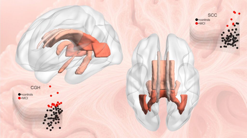
The researchers focused on four widely used measures of white matter microstructure across 21 major brain regions. By modeling how these measures vary by age and sex, they generated lifespan curves and percentile ranges that show what is typical at different stages of life.
![Statistical charts compiled from a large population allow brain abnormalities to be detected in new individuals. [Stevens INI]](https://www.genengnews.com/wp-content/uploads/2026/06/Julio-Villalon-press-release-image-1920x1080-1-1024x576.jpg)
The results revealed that white matter follows distinct developmental and aging trajectories, with some measures reaching peak maturity in early adulthood and others later in midlife.
“Brain development and brain aging are not uniform processes,” continued Villalón-Reina. “The brain’s neural pathways mature on distinct timelines, and some are more vulnerable to decline than others. Our model reveals this structure by merging data on a truly global scale.”
The scientists also discovered evidence for a longstanding theory of brain aging, sometimes described as last in, first out. According to this theory, brain pathways that develop last in childhood and adolescence tend to be more susceptible to decline in older age. The researchers observed that white matter regions that mature later did indeed decline faster in old age, offering new insight linking brain development and aging.
To demonstrate the model’s practical value, the researchers applied it to clinical datasets from people with mild cognitive impairment, dementia, and 22q11.2 deletion syndrome, a genetic condition that increases risk of schizophrenia.
In each case, the model identified alterations in the brain’s circuitry that deviated from age-expected norms. Importantly, these deviations were not identical across individuals with the same diagnosis, highlighting the value of a person-specific approach.

“This monumental study took seven years to complete,” explained Paul M. Thompson, PhD, associate director of the Stevens INI and senior author of the study. “The vast scale of the data and the fine scale of the brain features assessed means we can now evaluate your neural pathways relative to other people of the same age, sex, and demographics. We can see how your brain differs from what we would expect for a person of your age and sex, giving us a tool to use in clinical trials of treatments for dozens of brain diseases.”
When applied to people with dementia and mild cognitive impairment, the model detected atypical white matter patterns in brain regions involved in memory and interregional communication. In people with 22q11.2 deletion syndrome, it identified deviations in multiple key neural pathways, helping researchers discover which brain systems develop differently.
The reference charts may also help researchers evaluate treatments by tracking whether a person’s white matter measures move closer to the expected range, or whether a treatment slows the shift away from healthy patterns over time. The charts will now be used to compare more than 30 brain diseases and conditions, offering a common framework for studying how different disorders emerge, progress, and respond to intervention.
The models are also a publicly available resource that can be extended as additional brain imaging data become available. The methods are now being used to study neurological, psychiatric, and neurodevelopmental disorders by providing a common reference standard for white matter microstructure across the lifespan.
“This study demonstrates the power of large-scale, international data sharing to create tools the entire research community can use,” pointed out Arthur W. Toga, PhD, director of the Stevens INI and provost professor at USC. “By establishing a lifespan framework for the brain’s communication pathways, this work opens new opportunities to detect subtle disease-related changes, compare conditions more rigorously, and move toward a more individualized understanding of brain health.”
The post Disease Detection Gets Boost from Keck’s New Brain Reference Map appeared first on GEN – Genetic Engineering and Biotechnology News.

You know when you are at the eye doctor getting an updated prescription, and suddenly the world snaps into sharper focus? Physicists at the University of California (UC), Berkeley, have now done something similar for electron microscopy. By introducing phase contrast into a cryo‑electron microscope, they have delivered dramatically sharper images of some of biology’s smallest and most elusive proteins.
The advance comes from a new laser phase plate (LPP), described in the paper “Laser phase plate improves structure determination of small proteins by cryo‑EM,” which was published recently in Science. Led by physicist Holger Mueller, PhD, of UC Berkeley and Lawrence Berkeley National Laboratory, the team demonstrated that a laser‑driven phase plate can overcome one of cryo‑EM’s most persistent limitations: poor contrast for small proteins.

Cryo‑EM has transformed structural biology over the past decade, earning a Nobel Prize in 2017 for enabling high‑resolution structures without crystallization. But despite its impact, the technique still struggles with proteins below ~70 kilodaltons—a size range that includes about 90% of the human proteome. “Because of signal-to-noise limitations, the majority of human and animal proteins are too small to be analyzed by these methods [cryo-EM and cryoelectron tomography]. The increase in signal-to-noise ratio provided by this laser phase plate is expected to overcome these important limitations.”
The new LPP begins to address that problem. The LPP uses an intense, continuous‑wave laser to shift the phase of the electron beam itself. This produces true phase contrast without dimming or destabilizing the beam. Mueller described the laser focus as “75 kilowatts focused to a few microns… That’s more powerful than what you use for welding. It has more power than a military laser. It builds up the brightest continuous laser focus ever.”
Installed in a custom Thermo Fisher Titan Krios, the LPP immediately improved the clarity and resolvability of small proteins, including hemoglobin, which sits at the lower limit of what today’s cryo‑EM instruments can handle. As the authors wrote in the abstract: “Here, we show that the laser phase plate (LPP)… enhances the resolution in single-particle reconstruction of small proteins by improving specimen-motion correction, recovery of information from the early frames, as well as particle visualization, 3D classification, and alignment.”

These improvements were achieved using standard defocus ranges and reconstruction workflows. “For the most challenging cases—small particles, bad specimens—the laser produces a very considerable advantage,” Mueller said.
The impact extends beyond single‑particle analysis. Cryo‑electron tomography (cryo‑ET), which assembles multiple angular views of a molecule or protein into a three-dimensional image, stands to benefit even more. “With cryo-ET, we’re looking at small, very complicated cellular material that’s incredibly crowded inside the cell,” said Bridget Carragher, PhD, founding technical director of imaging at Biohub. “It’s like a forest of trees, and you’re trying to find one leaf on one tree in there. Cryo-ET needs a dramatic step forward in contrast, so we can start to see what’s going on inside the cell. That’s what the laser phase plate promises to give us.”
Biohub is developing a dual‑laser version of the system, designed to reduce component wear and minimize aberrations. Meanwhile, Mueller’s team is pushing toward imaging proteins as small as 17 kilodaltons, a threshold that would open access to vast regions of the human proteome previously invisible to cryo‑EM.
“This technology is a step function change for biology,” said Stephani Otte, PhD, Biohub’s vice president of imaging science. “What was once invisible will become visible—and that changes everything about how we understand disease.”
“The bottom line is, if you have a large protein and a really good sample—a fresh one or one frozen without bubbles, for example—you may not need the phase plate to get a single, high-quality image. But for a small protein and a bad sample, laser-on is best,” Mueller said. “This could fill an enormous gap in our knowledge of protein structures that can’t be crystallized or are too small for today’s cryo-EM. And it will be revolutionary for cryo-ET.”
The post Laser‑Driven Phase Contrast Enhances Cryo‑EM Resolution of Small Proteins appeared first on GEN – Genetic Engineering and Biotechnology News.
Uncategorized
STAT+: Updated: Tracking RFK Jr.’s promises to remake health in America
Updated June 11, 2026
WASHINGTON — A pledge to “Make America Healthy Again” earned Robert F. Kennedy Jr. his job atop U.S. health agencies a year and some change ago. He’s now had the opportunity to turn his words into action, with mixed results.
“All one needs” to prove the health secretary’s attentiveness is to “review my unprecedented list of accomplishments on a wide range of issues, all of which I drove,” Kennedy posted on X on Wednesday in response to a journalist.

Can any of the new obesity medications in development stand out from the pack? Which company just broke records with its IPO? And will the Food and Drug Administration allow greater access to experimental peptides?
We discuss all that and more on this week’s episode of “The Readout LOUD,” STAT’s biotech podcast.


Illinois’ financial crisis could bring the state to a halt
The final 6 ‘Game of Thrones’ episodes might feel like a full season
New Season 8 Walking Dead trailer flashes forward in time
Mod turns ‘Counter-Strike’ into a ‘Tekken’ clone with fighting chickens
Meet Superman’s grandfather in new trailer for Krypton
Disney’s live-action Aladdin finally finds its stars

Laser‑Driven Phase Contrast Enhances Cryo‑EM Resolution of Small Proteins

STAT+: Updated: Tracking RFK Jr.’s promises to remake health in America

An obesity drug deep-dive, and peptides move mainstream

RFK Jr. claims his calendar is publicly available. We’ve been trying to get it for a year

Nonprofit buys experimental cancer drug to maintain patient access
Potential Cocaine Addiction Targets Identified Through Genetic Mapping in Rats
Illinois’ financial crisis could bring the state to a halt
The final 6 ‘Game of Thrones’ episodes might feel like a full season
New Season 8 Walking Dead trailer flashes forward in time
Mod turns ‘Counter-Strike’ into a ‘Tekken’ clone with fighting chickens
Meet Superman’s grandfather in new trailer for Krypton
Disney’s live-action Aladdin finally finds its stars
-
 Uncategorized9 years ago
These ’90s fashion trends are making a comeback in 2017
-
Uncategorized9 years ago
According to Dior Couture, this taboo fashion accessory is back
-
Endpoints News3 months ago
Novartis to pay $2B upfront to take next-gen PI3Kα inhibitor from Synnovation
-
Uncategorized9 years ago
Phillies’ Aaron Altherr makes mind-boggling barehanded play
-
Uncategorized9 years ago
Uber and Lyft are finally available in all of New York State
-
Contributors9 years ago
The final 6 ‘Game of Thrones’ episodes might feel like a full season
-
Uncategorized9 years ago
Steph Curry finally got the contract he deserves from the Warriors
-
Uncategorized9 years ago
The old and New Edition cast comes together to perform